 Movie
Movie Controller

[English] 日本語
 Yorodumi
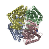
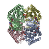
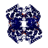
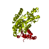
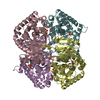
- PDB-6y91: Crystal structure of malate dehydrogenase from Plasmodium Falcipa... -
+ Open data

- Basic information
| Entry | Database: PDB / ID: 6y91 | ||||||
|---|---|---|---|---|---|---|---|
| Title | Crystal structure of malate dehydrogenase from Plasmodium Falciparum in complex with NADH | ||||||
 Components Components | Malate dehydrogenase | ||||||
 Keywords Keywords | OXIDOREDUCTASE | ||||||
| Function / homology |  Function and homology information Function and homology informationL-lactate dehydrogenase (NAD+) activity / lactate metabolic process / nucleotide binding Similarity search - Function | ||||||
| Biological species |  | ||||||
| Method |  X-RAY DIFFRACTION / MOLECULAR REPLACEMENT / Resolution: 2.5 Å X-RAY DIFFRACTION / MOLECULAR REPLACEMENT / Resolution: 2.5 Å | ||||||
 Authors Authors | Romero, A.R. / Calderone, V. / Gentili, M. / Lunev, S. / Groves, M. / Popowicz, G. / Domling, A. / Sattler, M. | ||||||
| Funding support |  Netherlands, 1items Netherlands, 1items
| ||||||
 Citation Citation | Journal: Commun Biol / Year: 2021 Title: A fragment-based approach identifies an allosteric pocket that impacts malate dehydrogenase activity. Authors: Reyes Romero, A. / Lunev, S. / Popowicz, G.M. / Calderone, V. / Gentili, M. / Sattler, M. / Plewka, J. / Taube, M. / Kozak, M. / Holak, T.A. / Domling, A.S.S. / Groves, M.R. #1: Journal: PLoS ONE / Year: 2018Title: Oligomeric interfaces as a tool in drug discovery: Specific interference with activity of malate dehydrogenase of Plasmodium falciparum in vitro. Authors: Lunev, S. / Butzloff, S. / Romero, A.R. / Linzke, M. / Batista, F.A. / Meissner, K.A. / Muller, I.B. / Adawy, A. / Wrenger, C. / Groves, M.R. | ||||||
| History |
|
- Structure visualization
| Structure viewer | Molecule: MolmilJmol/JSmol |
|---|
- Downloads & links
-Download
| PDBx/mmCIF format | 6y91.cif.gz | 466.4 KB | Display | PDBx/mmCIF format |
|---|---|---|---|---|
| PDB format | pdb6y91.ent.gz | 383.7 KB | Display | PDB format |
| PDBx/mmJSON format | 6y91.json.gz | Tree view | PDBx/mmJSON format | |
| Others |  Other downloads Other downloads |
-Validation report
| Arichive directory | https://data.pdbj.org/pub/pdb/validation_reports/y9/6y91ftp://data.pdbj.org/pub/pdb/validation_reports/y9/6y91 | HTTPS FTP |
|---|
-Related structure data
| Related structure data |  5nfrS S: Starting model for refinement |
|---|---|
| Similar structure data |


-Links
 PDBj
PDBj


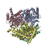
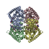
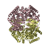
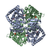
- Assembly
| Deposited unit | 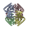
| ||||||||
|---|---|---|---|---|---|---|---|---|---|
| 1 |
| ||||||||
| Unit cell |
|
-Components
| #1: Protein | Mass: 35362.922 Da / Num. of mol.: 4 Source method: isolated from a genetically manipulated source Source: (gene. exp.) Production host:  References: UniProt: Q6VVP7, (S)-malate dehydrogenase (NAD+, oxaloacetate-forming) #2: Chemical | ChemComp-NAD /   Mass: 663.425 Da / Num. of mol.: 4 / Source method: obtained synthetically / Formula: C21H27N7O14P2 / Feature type: SUBJECT OF INVESTIGATION / Comment: NAD*YM Mass: 663.425 Da / Num. of mol.: 4 / Source method: obtained synthetically / Formula: C21H27N7O14P2 / Feature type: SUBJECT OF INVESTIGATION / Comment: NAD*YM#3: Water | ChemComp-HOH / |  Mass: 18.015 Da / Num. of mol.: 143 / Source method: isolated from a natural source / Formula: H2O Mass: 18.015 Da / Num. of mol.: 143 / Source method: isolated from a natural source / Formula: H2OHas ligand of interest | Y | |
|---|
-Experimental details
-Experiment
| Experiment | Method: X-RAY DIFFRACTION / Number of used crystals: 1 |
|---|
- Sample preparation
| Crystal | Density Matthews: 2.52 Å3/Da / Density % sol: 51.28 % |
|---|---|
| Crystal grow | Temperature: 293 K / Method: vapor diffusion, sitting drop / pH: 7.5 / Details: 1.4 M trisodium citrate, 0.1 M Hepes, 10 mM NADH |
-Data collection
| Diffraction | Mean temperature: 100 K / Serial crystal experiment: N |
|---|---|
| Diffraction source | Source: SEALED TUBE / Type: BRUKER D8 QUEST / Wavelength: 1.54 Å |
| Detector | Type: Bruker PHOTON II / Detector: PIXEL / Date: Mar 16, 2019 |
| Radiation | Monochromator: mirrors / Protocol: SINGLE WAVELENGTH / Monochromatic (M) / Laue (L): M / Scattering type: x-ray |
| Radiation wavelength | Wavelength: 1.54 Å / Relative weight: 1 |
| Reflection | Resolution: 2.5→18.27 Å / Num. obs: 46208 / % possible obs: 99.7 % / Observed criterion σ(F): 0 / Observed criterion σ(I): 0 / Redundancy: 16.8 % / CC1/2: 0.99 / Rmerge(I) obs: 0.2 / Net I/σ(I): 9.3 |
| Reflection shell | Resolution: 2.5→2.57 Å / Redundancy: 16.3 % / Rmerge(I) obs: 1.1 / Mean I/σ(I) obs: 2.1 / Num. unique obs: 3249 / CC1/2: 0.61 / % possible all: 96.6 |
- Processing
| Software |
| |||||||||||||||||||||||||||||||||||||||||||||||||||||||||||||||||||||||||||||||||||||||||||||||||||||||||||||||||||||||||||||
|---|---|---|---|---|---|---|---|---|---|---|---|---|---|---|---|---|---|---|---|---|---|---|---|---|---|---|---|---|---|---|---|---|---|---|---|---|---|---|---|---|---|---|---|---|---|---|---|---|---|---|---|---|---|---|---|---|---|---|---|---|---|---|---|---|---|---|---|---|---|---|---|---|---|---|---|---|---|---|---|---|---|---|---|---|---|---|---|---|---|---|---|---|---|---|---|---|---|---|---|---|---|---|---|---|---|---|---|---|---|---|---|---|---|---|---|---|---|---|---|---|---|---|---|---|---|---|
| Refinement | Method to determine structure: MOLECULAR REPLACEMENT Starting model: 5nfr Resolution: 2.5→18.27 Å / SU ML: 0.38 / Cross valid method: FREE R-VALUE / σ(F): 1.36 / Phase error: 28
| |||||||||||||||||||||||||||||||||||||||||||||||||||||||||||||||||||||||||||||||||||||||||||||||||||||||||||||||||||||||||||||
| Solvent computation | Shrinkage radii: 0.9 Å / VDW probe radii: 1.11 Å | |||||||||||||||||||||||||||||||||||||||||||||||||||||||||||||||||||||||||||||||||||||||||||||||||||||||||||||||||||||||||||||
| Refinement step | Cycle: LAST / Resolution: 2.5→18.27 Å
| |||||||||||||||||||||||||||||||||||||||||||||||||||||||||||||||||||||||||||||||||||||||||||||||||||||||||||||||||||||||||||||
| Refine LS restraints |
| |||||||||||||||||||||||||||||||||||||||||||||||||||||||||||||||||||||||||||||||||||||||||||||||||||||||||||||||||||||||||||||
| LS refinement shell |
| |||||||||||||||||||||||||||||||||||||||||||||||||||||||||||||||||||||||||||||||||||||||||||||||||||||||||||||||||||||||||||||
| Refinement TLS params. | Method: refined / Refine-ID: X-RAY DIFFRACTION
| |||||||||||||||||||||||||||||||||||||||||||||||||||||||||||||||||||||||||||||||||||||||||||||||||||||||||||||||||||||||||||||
| Refinement TLS group |
|
