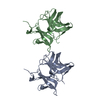
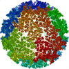
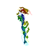
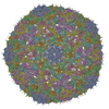
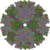
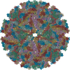
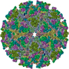
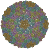
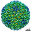
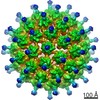
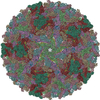
ジャーナル: J Virol / 年: 2011 タイトル: The structure of barmah forest virus as revealed by cryo-electron microscopy at a 6-angstrom resolution has detailed transmembrane protein architecture and interactions. 著者: Victor A Kostyuchenko / Joanita Jakana / Xiangan Liu / Andrew D Haddow / Myint Aung / Scott C Weaver / Wah Chiu / Shee-Mei Lok / 要旨: Barmah Forest virus (BFV) is a mosquito-borne alphavirus that infects humans. A 6-Å-resolution cryo-electron microscopy three-dimensional structure of BFV exhibits a typical alphavirus organization, ...Barmah Forest virus (BFV) is a mosquito-borne alphavirus that infects humans. A 6-Å-resolution cryo-electron microscopy three-dimensional structure of BFV exhibits a typical alphavirus organization, with RNA-containing nucleocapsid surrounded by a bilipid membrane anchored with the surface proteins E1 and E2. The map allows details of the transmembrane regions of E1 and E2 to be seen. The C-terminal end of the E2 transmembrane helix binds to the capsid protein. Following the E2 transmembrane helix, a short α-helical endodomain lies on the inner surface of the lipid envelope. The E2 endodomain interacts with E1 transmembrane helix from a neighboring E1-E2 trimeric spike, thereby acting as a spacer and a linker between spikes. In agreement with previous mutagenesis studies, the endodomain plays an important role in recruiting other E1-E2 spikes to the budding site during virus assembly. The E2 endodomain may thus serve as a target for antiviral drug design.
履歴
登録
2011年3月31日
登録サイト: PDBE / 処理サイト: PDBE
改定 1.0
2012年4月18日
Provider: repository / タイプ: Initial release
改定 1.1
2013年3月20日
Group: Other
改定 1.2
2017年8月23日
Group: Data collection / カテゴリ: em_software Item: _em_software.fitting_id / _em_software.image_processing_id
改定 1.3
2019年10月23日
Group: Data collection / Other / カテゴリ: cell / Item: _cell.Z_PDB
モード: BRIGHT FIELD / 倍率(公称値): 50000 X / 最大 デフォーカス(公称値): 3500 nm / 最小 デフォーカス(公称値): 500 nm / Cs: 4.1 mm
試料ホルダ
温度: 100 K
撮影
電子線照射量: 20 e/Å2 / フィルム・検出器のモデル: GENERIC GATAN
画像スキャン
デジタル画像の数: 760
放射波長
相対比: 1
-
解析
EMソフトウェア
ID
名称
カテゴリ
1
UCSF Chimera
モデルフィッティング
2
EMAN
3次元再構成
3
MPSA
3次元再構成
CTF補正
詳細: INDIVIDUAL PARTICLES
対称性
点対称性: I (正20面体型対称)
3次元再構成
手法: CROSS-COMMON LINES, MULTIPATH SIMULTANEOUS ANNEALING PROTOCOL 解像度: 5 Å / 粒子像の数: 5169 / ピクセルサイズ(公称値): 1.42 Å / ピクセルサイズ(実測値): 1.42 Å 詳細: THE MODELS WERE BUILD USING MODELLER FOR HOMOLOGY -BASED MODELLING AND USING VMD WITH NAMD FOR FLEXIBLE FITTING INTO CRYO-EM DENSITY. CHAIN A DOES NOT CONTAIN RNA-BINDING PART, ABOUT 80 N- ...詳細: THE MODELS WERE BUILD USING MODELLER FOR HOMOLOGY -BASED MODELLING AND USING VMD WITH NAMD FOR FLEXIBLE FITTING INTO CRYO-EM DENSITY. CHAIN A DOES NOT CONTAIN RNA-BINDING PART, ABOUT 80 N-TERMINAL RESIDUES. THE STRUCTURE WAS MODELED BASED ON HOMOLOGY TO PROTEIN WITH KNOWN STURUCTURE AND CRYO-EM DENSITY FITTING. SUBMISSION BASED ON EXPERIMENTAL DATA FROM EMDB EMD- 1886. (DEPOSITION ID: 7893). 対称性のタイプ: POINT
原子モデル構築
プロトコル: RIGID BODY FIT / 空間: REAL / 詳細: REFINEMENT PROTOCOL--RIGID BODY
 ムービー
ムービー コントローラー


 データを開く
 基本情報
基本情報 要素
要素 キーワード
キーワード 機能・相同性情報
 BARMAH FOREST VIRUS (ウイルス)
 データ登録者
 引用
引用
 構造の表示
構造の表示 ダウンロードとリンク
 その他のダウンロード

 PDBj
PDBj


 集合体
集合体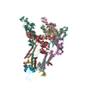


 MESOCRICETUS AURATUS (ネズミ) / 参照: UniProt: P89946, togavirin
 試料調製
試料調製 電子顕微鏡撮影
 FIELD EMISSION GUN / 加速電圧: 300 kV / 照射モード: FLOOD BEAM
 解析
解析