 Movie
Movie Controller

[English] 日本語
 Yorodumi
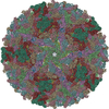
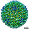
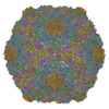
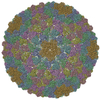
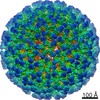
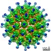
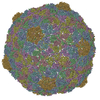
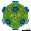
- PDB-6c21: Capsid protein in the Staphylococcus aureus phage 80alpha mature ... -
+ Open data

- Basic information
| Entry | Database: PDB / ID: 6c21 | ||||||
|---|---|---|---|---|---|---|---|
| Title | Capsid protein in the Staphylococcus aureus phage 80alpha mature capsid | ||||||
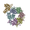
 Components Components | Major head protein | ||||||
 Keywords Keywords | VIRUS / major capsid protein / HK97-like fold / mature capsid | ||||||
| Function / homology | : / Phage capsid / Phage capsid family / viral capsid / Major capsid protein Function and homology information Function and homology information | ||||||
| Biological species |  Staphylococcus virus 80alpha Staphylococcus virus 80alpha | ||||||
| Method | ELECTRON MICROSCOPY / single particle reconstruction / cryo EM / Resolution: 5.2 Å | ||||||
 Authors Authors | Kizziah, J.L. / Dearborn, A.D. / Dokland, T. | ||||||
| Funding support |  United States, 1items United States, 1items
| ||||||
 Citation Citation | Journal: Viruses / Year: 2017 Title: Cleavage and Structural Transitions during Maturation of Staphylococcus aureus Bacteriophage 80α and SaPI1 Capsids. Authors: James L Kizziah / Keith A Manning / Altaira D Dearborn / Erin A Wall / Laura Klenow / Rosanne L L Hill / Michael S Spilman / Scott M Stagg / Gail E Christie / Terje Dokland / Abstract: In the tailed bacteriophages, DNA is packaged into spherical procapsids, leading to expansion into angular, thin-walled mature capsids. In many cases, this maturation is accompanied by cleavage of ...In the tailed bacteriophages, DNA is packaged into spherical procapsids, leading to expansion into angular, thin-walled mature capsids. In many cases, this maturation is accompanied by cleavage of the major capsid protein (CP) and other capsid-associated proteins, including the scaffolding protein (SP) that serves as a chaperone for the assembly process. bacteriophage 80α is capable of high frequency mobilization of mobile genetic elements called pathogenicity islands (SaPIs), such as SaPI1. SaPI1 redirects the assembly pathway of 80α to form capsids that are smaller than those normally made by the phage alone. Both CP and SP of 80α are N-terminally processed by a host-encoded protease, Prp. We have analyzed phage mutants that express pre-cleaved or uncleavable versions of CP or SP, and show that the N-terminal sequence in SP is absolutely required for assembly, but does not need to be cleaved in order to produce viable capsids. Mutants with pre-cleaved or uncleavable CP display normal viability. We have used cryo-EM to solve the structures of mature capsids from an 80α mutant expressing uncleavable CP, and from wildtype SaPI1. Comparisons with structures of 80α and SaPI1 procapsids show that capsid maturation involves major conformational changes in CP, consistent with a release of the CP N-arm by SP. The hexamers reorganize during maturation to accommodate the different environments in the 80α and SaPI1 capsids. | ||||||
| History |
|
- Structure visualization
| Movie |
Movie viewer |
|---|---|
| Structure viewer | Molecule: MolmilJmol/JSmol |
- Downloads & links
-Download
| PDBx/mmCIF format | 6c21.cif.gz | 301.6 KB | Display | PDBx/mmCIF format |
|---|---|---|---|---|
| PDB format | pdb6c21.ent.gz | 208.6 KB | Display | PDB format |
| PDBx/mmJSON format | 6c21.json.gz | Tree view | PDBx/mmJSON format | |
| Others |  Other downloads Other downloads |
-Validation report
| Arichive directory | https://data.pdbj.org/pub/pdb/validation_reports/c2/6c21ftp://data.pdbj.org/pub/pdb/validation_reports/c2/6c21 | HTTPS FTP |
|---|
-Related structure data
| Related structure data | 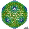 7332MC 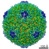 7333C 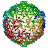 6c22C M: map data used to model this data C: citing same article ( |
|---|---|
| Similar structure data |

-Links
 PDBj
PDBj

- Assembly
| Deposited unit | 
|
|---|---|
| 1 | x 60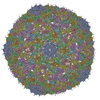
|
| 2 |
|
| 3 | x 5
|
| 4 | x 6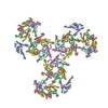
|
| 5 | 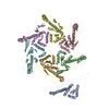
|
| Symmetry | Point symmetry: (Schoenflies symbol: I (icosahedral)) |
-Components
| #1: Protein | Mass: 36846.883 Da / Num. of mol.: 7 Source method: isolated from a genetically manipulated source Source: (gene. exp.) Staphylococcus virus 80alpha / Cell line (production host): RN450 / Production host:   Staphylococcus aureus (bacteria) / Strain (production host): ST208 / References: UniProt: A4ZFB3 Staphylococcus aureus (bacteria) / Strain (production host): ST208 / References: UniProt: A4ZFB3 |
|---|
-Experimental details
-Experiment
| Experiment | Method: ELECTRON MICROSCOPY |
|---|---|
| EM experiment | Aggregation state: PARTICLE / 3D reconstruction method: single particle reconstruction |
- Sample preparation
| Component | Name: Staphylococcus phage 80alpha / Type: VIRUS / Entity ID: all / Source: RECOMBINANT |
|---|---|
| Molecular weight | Value: 15.46 MDa / Experimental value: NO |
| Source (natural) | Organism: Staphylococcus phage 80alpha (virus) |
| Source (recombinant) | Organism: Staphylococcus aureus (bacteria) |
| Details of virus | Empty: YES / Enveloped: NO / Isolate: SPECIES / Type: VIRION |
| Natural host | Organism: Staphylococcus aureus |
| Virus shell | Name: Capsid / Diameter: 630 nm / Triangulation number (T number): 7 |
| Buffer solution | pH: 8 |
| Specimen | Embedding applied: NO / Shadowing applied: NO / Staining applied: NO / Vitrification applied: YES |
| Vitrification | Cryogen name: ETHANE |
- Electron microscopy imaging
| Experimental equipment |  Model: Titan Krios / Image courtesy: FEI Company |
|---|---|
| Microscopy | Model: FEI TITAN KRIOS |
| Electron gun | Electron source:  FIELD EMISSION GUN / Accelerating voltage: 120 kV / Illumination mode: FLOOD BEAM FIELD EMISSION GUN / Accelerating voltage: 120 kV / Illumination mode: FLOOD BEAM |
| Electron lens | Mode: BRIGHT FIELD / Nominal magnification: 96000 X / Nominal defocus max: 3000 nm / Nominal defocus min: 1250 nm |
| Image recording | Electron dose: 15 e/Å2 / Film or detector model: GATAN ULTRASCAN 4000 (4k x 4k) |
- Processing
| Software | Name: REFMAC / Version: 5.8.0088 / Classification: refinement | ||||||||||||||||||||||||||||||||||||||||||||||||||||||||||||||||||||||||||||||||||||||||||||||||||||||||||
|---|---|---|---|---|---|---|---|---|---|---|---|---|---|---|---|---|---|---|---|---|---|---|---|---|---|---|---|---|---|---|---|---|---|---|---|---|---|---|---|---|---|---|---|---|---|---|---|---|---|---|---|---|---|---|---|---|---|---|---|---|---|---|---|---|---|---|---|---|---|---|---|---|---|---|---|---|---|---|---|---|---|---|---|---|---|---|---|---|---|---|---|---|---|---|---|---|---|---|---|---|---|---|---|---|---|---|---|
| EM software |
| ||||||||||||||||||||||||||||||||||||||||||||||||||||||||||||||||||||||||||||||||||||||||||||||||||||||||||
| CTF correction | Details: Estimated with ACE software / Type: PHASE FLIPPING ONLY | ||||||||||||||||||||||||||||||||||||||||||||||||||||||||||||||||||||||||||||||||||||||||||||||||||||||||||
| Particle selection | Num. of particles selected: 17946 | ||||||||||||||||||||||||||||||||||||||||||||||||||||||||||||||||||||||||||||||||||||||||||||||||||||||||||
| Symmetry | Point symmetry: I (icosahedral) | ||||||||||||||||||||||||||||||||||||||||||||||||||||||||||||||||||||||||||||||||||||||||||||||||||||||||||
| 3D reconstruction | Resolution: 5.2 Å / Resolution method: FSC 0.143 CUT-OFF / Num. of particles: 11843 / Symmetry type: POINT | ||||||||||||||||||||||||||||||||||||||||||||||||||||||||||||||||||||||||||||||||||||||||||||||||||||||||||
| Atomic model building | Protocol: RIGID BODY FIT / Space: REAL | ||||||||||||||||||||||||||||||||||||||||||||||||||||||||||||||||||||||||||||||||||||||||||||||||||||||||||
| Refinement | Resolution: 5.2→168 Å / Cor.coef. Fo:Fc: 0.845 / SU B: 93.003 / SU ML: 0.938 / ESU R: 1.701 Stereochemistry target values: MAXIMUM LIKELIHOOD WITH PHASES Details: HYDROGENS HAVE BEEN USED IF PRESENT IN THE INPUT
| ||||||||||||||||||||||||||||||||||||||||||||||||||||||||||||||||||||||||||||||||||||||||||||||||||||||||||
| Solvent computation | Ion probe radii: 0.8 Å / Shrinkage radii: 0.8 Å / VDW probe radii: 1.2 Å / Solvent model: MASK | ||||||||||||||||||||||||||||||||||||||||||||||||||||||||||||||||||||||||||||||||||||||||||||||||||||||||||
| Displacement parameters | Biso mean: 176.424 Å2
| ||||||||||||||||||||||||||||||||||||||||||||||||||||||||||||||||||||||||||||||||||||||||||||||||||||||||||
| Refine LS restraints |
|