Movie
Movie Controller


+ Open data

- Basic information
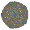
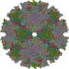
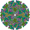
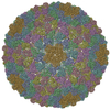
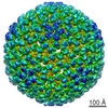
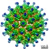
| Entry | Database: PDB / ID: 2yew | ||||||
|---|---|---|---|---|---|---|---|
| Title | Modeling Barmah Forest virus structural proteins | ||||||
 Components Components |
| ||||||
 Keywords Keywords | VIRUS / ALPHAVIRUS / MOLECULAR DYNAMICS | ||||||
| Function / homology |  Function and homology information Function and homology informationtogavirin / T=4 icosahedral viral capsid / symbiont-mediated suppression of host toll-like receptor signaling pathway / host cell cytoplasm / serine-type endopeptidase activity / fusion of virus membrane with host endosome membrane / symbiont entry into host cell / virion attachment to host cell / host cell nucleus / host cell plasma membrane ...togavirin / T=4 icosahedral viral capsid / symbiont-mediated suppression of host toll-like receptor signaling pathway / host cell cytoplasm / serine-type endopeptidase activity / fusion of virus membrane with host endosome membrane / symbiont entry into host cell / virion attachment to host cell / host cell nucleus / host cell plasma membrane / virion membrane / structural molecule activity / proteolysis / RNA binding / membrane Similarity search - Function | ||||||
| Biological species |  BARMAH FOREST VIRUS BARMAH FOREST VIRUS | ||||||
| Method | ELECTRON MICROSCOPY / single particle reconstruction / cryo EM / Resolution: 5 Å | ||||||
 Authors Authors | Kostyuchenko, V.A. / Jakana, J. / Liu, X. / Haddow, A.D. / Aung, M. / Weaver, S.C. / Chiu, W. / Lok, S.M. | ||||||
 Citation Citation | Journal: J Virol / Year: 2011 Title: The structure of barmah forest virus as revealed by cryo-electron microscopy at a 6-angstrom resolution has detailed transmembrane protein architecture and interactions. Authors: Victor A Kostyuchenko / Joanita Jakana / Xiangan Liu / Andrew D Haddow / Myint Aung / Scott C Weaver / Wah Chiu / Shee-Mei Lok /  Abstract: Barmah Forest virus (BFV) is a mosquito-borne alphavirus that infects humans. A 6-Å-resolution cryo-electron microscopy three-dimensional structure of BFV exhibits a typical alphavirus organization, ...Barmah Forest virus (BFV) is a mosquito-borne alphavirus that infects humans. A 6-Å-resolution cryo-electron microscopy three-dimensional structure of BFV exhibits a typical alphavirus organization, with RNA-containing nucleocapsid surrounded by a bilipid membrane anchored with the surface proteins E1 and E2. The map allows details of the transmembrane regions of E1 and E2 to be seen. The C-terminal end of the E2 transmembrane helix binds to the capsid protein. Following the E2 transmembrane helix, a short α-helical endodomain lies on the inner surface of the lipid envelope. The E2 endodomain interacts with E1 transmembrane helix from a neighboring E1-E2 trimeric spike, thereby acting as a spacer and a linker between spikes. In agreement with previous mutagenesis studies, the endodomain plays an important role in recruiting other E1-E2 spikes to the budding site during virus assembly. The E2 endodomain may thus serve as a target for antiviral drug design. | ||||||
| History |
|
- Structure visualization
| Movie |
Movie viewer |
|---|---|
| Structure viewer | Molecule: MolmilJmol/JSmol |
- Downloads & links
-Download
| PDBx/mmCIF format | 2yew.cif.gz | 697.2 KB | Display | PDBx/mmCIF format |
|---|---|---|---|---|
| PDB format | pdb2yew.ent.gz | 536.4 KB | Display | PDB format |
| PDBx/mmJSON format | 2yew.json.gz | Tree view | PDBx/mmJSON format | |
| Others |  Other downloads Other downloads |
-Validation report
| Summary document | 2yew_validation.pdf.gz | 1.3 MB | Display | wwPDB validaton report |
|---|---|---|---|---|
| Full document | 2yew_full_validation.pdf.gz | 1.5 MB | Display | |
| Data in XML | 2yew_validation.xml.gz | 125.8 KB | Display | |
| Data in CIF | 2yew_validation.cif.gz | 183.2 KB | Display | |
| Arichive directory | https://data.pdbj.org/pub/pdb/validation_reports/ye/2yewftp://data.pdbj.org/pub/pdb/validation_reports/ye/2yew | HTTPS FTP |
-Related structure data
| Related structure data |  1886MC M: map data used to model this data C: citing same article ( |
|---|---|
| Similar structure data |

-Links
 PDBj
PDBj


- Assembly
| Deposited unit | 
|
|---|---|
| 1 | x 60
|
| 2 |
|
| 3 | x 5
|
| 4 | x 6
|
| 5 | 
|
| Symmetry | Point symmetry: (Schoenflies symbol: I (icosahedral)) |
-Components
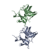
| #1: Protein | Mass: 28311.041 Da / Num. of mol.: 4 Source method: isolated from a genetically manipulated source Source: (gene. exp.) BARMAH FOREST VIRUS / Cell line (production host): BHK-21 / Production host:  MESOCRICETUS AURATUS (golden hamster) / References: UniProt: P89946, togavirin MESOCRICETUS AURATUS (golden hamster) / References: UniProt: P89946, togavirin#2: Protein | Mass: 46283.312 Da / Num. of mol.: 4 Source method: isolated from a genetically manipulated source Source: (gene. exp.) BARMAH FOREST VIRUS / Cell line (production host): BHK-21 / Production host: MESOCRICETUS AURATUS (golden hamster) / References: UniProt: P89946#3: Protein | Mass: 46185.648 Da / Num. of mol.: 4 Source method: isolated from a genetically manipulated source Source: (gene. exp.) BARMAH FOREST VIRUS / Cell line (production host): BHK-21 / Production host: MESOCRICETUS AURATUS (golden hamster) / References: UniProt: P89946Has protein modification | Y | |
|---|
-Experimental details
-Experiment
| Experiment | Method: ELECTRON MICROSCOPY |
|---|---|
| EM experiment | Aggregation state: PARTICLE / 3D reconstruction method: single particle reconstruction |
- Sample preparation
| Component | Name: BARMAH FOREST VIRUS / Type: VIRUS |
|---|---|
| Buffer solution | pH: 7.4 / Details: 0.05M Tris-HCl, pH 7.4, 0.1M NaCl, 0.001M EDTA |
| Specimen | Embedding applied: NO / Shadowing applied: NO / Staining applied: NO / Vitrification applied: YES |
| Specimen support | Details: HOLEY CARBON |
| Vitrification | Instrument: FEI VITROBOT MARK I / Cryogen name: ETHANE |
- Electron microscopy imaging
| Microscopy | Model: JEOL 3200FSC / Date: Jan 10, 2010 |
|---|---|
| Electron gun | Electron source:  FIELD EMISSION GUN / Accelerating voltage: 300 kV / Illumination mode: FLOOD BEAM FIELD EMISSION GUN / Accelerating voltage: 300 kV / Illumination mode: FLOOD BEAM |
| Electron lens | Mode: BRIGHT FIELD / Nominal magnification: 50000 X / Nominal defocus max: 3500 nm / Nominal defocus min: 500 nm / Cs: 4.1 mm |
| Specimen holder | Temperature: 100 K |
| Image recording | Electron dose: 20 e/Å2 / Film or detector model: GENERIC GATAN |
| Image scans | Num. digital images: 760 |
| Radiation wavelength | Relative weight: 1 |
- Processing
| EM software |
| ||||||||||||||||||||||||||||
|---|---|---|---|---|---|---|---|---|---|---|---|---|---|---|---|---|---|---|---|---|---|---|---|---|---|---|---|---|---|
| CTF correction | Details: INDIVIDUAL PARTICLES | ||||||||||||||||||||||||||||
| Symmetry | Point symmetry: I (icosahedral) | ||||||||||||||||||||||||||||
| 3D reconstruction | Method: CROSS-COMMON LINES, MULTIPATH SIMULTANEOUS ANNEALING PROTOCOL Resolution: 5 Å / Num. of particles: 5169 / Nominal pixel size: 1.42 Å / Actual pixel size: 1.42 Å Details: THE MODELS WERE BUILD USING MODELLER FOR HOMOLOGY -BASED MODELLING AND USING VMD WITH NAMD FOR FLEXIBLE FITTING INTO CRYO-EM DENSITY. CHAIN A DOES NOT CONTAIN RNA-BINDING PART, ABOUT 80 N- ...Details: THE MODELS WERE BUILD USING MODELLER FOR HOMOLOGY -BASED MODELLING AND USING VMD WITH NAMD FOR FLEXIBLE FITTING INTO CRYO-EM DENSITY. CHAIN A DOES NOT CONTAIN RNA-BINDING PART, ABOUT 80 N-TERMINAL RESIDUES. THE STRUCTURE WAS MODELED BASED ON HOMOLOGY TO PROTEIN WITH KNOWN STURUCTURE AND CRYO-EM DENSITY FITTING. SUBMISSION BASED ON EXPERIMENTAL DATA FROM EMDB EMD- 1886. (DEPOSITION ID: 7893). Symmetry type: POINT | ||||||||||||||||||||||||||||
| Atomic model building | Protocol: RIGID BODY FIT / Space: REAL / Details: REFINEMENT PROTOCOL--RIGID BODY | ||||||||||||||||||||||||||||
| Atomic model building |
| ||||||||||||||||||||||||||||
| Refinement | Highest resolution: 5 Å | ||||||||||||||||||||||||||||
| Refinement step | Cycle: LAST / Highest resolution: 5 Å
|