 Movie
Movie Controller

[English] 日本語
 Yorodumi
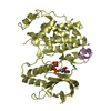
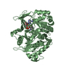
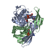
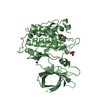
- PDB-2y9q: Crystal structure of human ERK2 complexed with a MAPK docking peptide -
+ Open data

- Basic information
| Entry | Database: PDB / ID: 2y9q | ||||||
|---|---|---|---|---|---|---|---|
| Title | Crystal structure of human ERK2 complexed with a MAPK docking peptide | ||||||
 Components Components |
| ||||||
 Keywords Keywords | TRANSFERASE / SIGNALING / PROTEIN-PROTEIN INTERACTION | ||||||
| Function / homology |  Function and homology information Function and homology informationcalcium-dependent protein serine/threonine kinase activity / phospho-PLA2 pathway / Signaling by MAPK mutants / RAF-independent MAPK1/3 activation / Suppression of apoptosis / Gastrin-CREB signalling pathway via PKC and MAPK / Signaling by Activin / cytosine metabolic process / cardiac neural crest cell development involved in heart development / interleukin-34-mediated signaling pathway ...calcium-dependent protein serine/threonine kinase activity / phospho-PLA2 pathway / Signaling by MAPK mutants / RAF-independent MAPK1/3 activation / Suppression of apoptosis / Gastrin-CREB signalling pathway via PKC and MAPK / Signaling by Activin / cytosine metabolic process / cardiac neural crest cell development involved in heart development / interleukin-34-mediated signaling pathway / caveolin-mediated endocytosis / outer ear morphogenesis / trachea formation / response to epidermal growth factor / labyrinthine layer blood vessel development / Signaling by NODAL / Signaling by MAP2K mutants / RSK activation / ERKs are inactivated / Bergmann glial cell differentiation / lung morphogenesis / calcium/calmodulin-dependent protein kinase activity / Regulation of the apoptosome activity / Golgi Cisternae Pericentriolar Stack Reorganization / positive regulation of macrophage proliferation / : / mammary gland epithelial cell proliferation / regulation of Golgi inheritance / Signaling by LTK in cancer / positive regulation of peptidyl-threonine phosphorylation / face development / ERBB signaling pathway / regulation of early endosome to late endosome transport / thyroid gland development / Negative feedback regulation of MAPK pathway / regulation of stress-activated MAPK cascade / positive regulation of neuroinflammatory response / IFNG signaling activates MAPKs / Frs2-mediated activation / response to exogenous dsRNA / Activation of the AP-1 family of transcription factors / ERBB2-ERBB3 signaling pathway / ERK/MAPK targets / RUNX2 regulates osteoblast differentiation / regulation of cytoskeleton organization / positive regulation of macrophage chemotaxis / MAPK1 (ERK2) activation / Recycling pathway of L1 / pseudopodium / positive regulation of telomere maintenance / regulation of ossification / MAP kinase activity / Advanced glycosylation endproduct receptor signaling / mitogen-activated protein kinase / Regulation of HSF1-mediated heat shock response / Estrogen-dependent nuclear events downstream of ESR-membrane signaling / stress-activated MAPK cascade / RHO GTPases Activate NADPH Oxidases / negative regulation of cell differentiation / Signal attenuation / ERK1 and ERK2 cascade / RHO GTPases Activate WASPs and WAVEs / Growth hormone receptor signaling / Schwann cell development / thymus development / phosphatase binding / Estrogen-stimulated signaling through PRKCZ / myelination / NPAS4 regulates expression of target genes / lipopolysaccharide-mediated signaling pathway / phosphotyrosine residue binding / ciliary tip / insulin-like growth factor receptor signaling pathway / Nuclear events stimulated by ALK signaling in cancer / RNA polymerase II CTD heptapeptide repeat kinase activity / Transcriptional and post-translational regulation of MITF-M expression and activity / ESR-mediated signaling / NCAM signaling for neurite out-growth / cellular response to amino acid starvation / B cell receptor signaling pathway / Regulation of PTEN gene transcription / Signal transduction by L1 / chemokine-mediated signaling pathway / response to nicotine / FCGR3A-mediated phagocytosis / FCERI mediated MAPK activation / Negative regulation of FGFR3 signaling / Negative regulation of FGFR2 signaling / Negative regulation of FGFR4 signaling / cellular response to tumor necrosis factor / Downregulation of SMAD2/3:SMAD4 transcriptional activity / Negative regulation of FGFR1 signaling / SMAD2/SMAD3:SMAD4 heterotrimer regulates transcription / Signaling by high-kinase activity BRAF mutants / Spry regulation of FGF signaling / positive regulation of cholesterol biosynthetic process / MAP2K and MAPK activation / Regulation of actin dynamics for phagocytic cup formation / Negative Regulation of CDH1 Gene Transcription / Oncogene Induced Senescence Similarity search - Function | ||||||
| Biological species |  HOMO SAPIENS (human) HOMO SAPIENS (human) | ||||||
| Method |  X-RAY DIFFRACTION / MOLECULAR REPLACEMENT / Resolution: 1.55 Å X-RAY DIFFRACTION / MOLECULAR REPLACEMENT / Resolution: 1.55 Å | ||||||
 Authors Authors | Barkai, T. / Garai, A. / Toeroe, I. / Remenyi, A. | ||||||
 Citation Citation | Journal: Sci. Signal / Year: 2012 Title: Specificity of Linear Motifs that Bind to a Common Mitogen-Activated Protein Kinase Docking Groove. Authors: Garai, A. / Zeke, A. / Gogl, G. / Toro, I. / Fordos, F. / Blankenburg, H. / Barkai, T. / Varga, J. / Alexa, A. / Emig, D. / Albrecht, M. / Remenyi, A. | ||||||
| History |
|
- Structure visualization
| Structure viewer | Molecule: MolmilJmol/JSmol |
|---|
- Downloads & links
-Download
| PDBx/mmCIF format | 2y9q.cif.gz | 177.1 KB | Display | PDBx/mmCIF format |
|---|---|---|---|---|
| PDB format | pdb2y9q.ent.gz | 140.4 KB | Display | PDB format |
| PDBx/mmJSON format | 2y9q.json.gz | Tree view | PDBx/mmJSON format | |
| Others |  Other downloads Other downloads |
-Validation report
| Arichive directory | https://data.pdbj.org/pub/pdb/validation_reports/y9/2y9qftp://data.pdbj.org/pub/pdb/validation_reports/y9/2y9q | HTTPS FTP |
|---|
-Related structure data
| Related structure data | 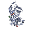 2xrwC  2xs0C  2y8oC  3teiC  4fmqC  2gphS S: Starting model for refinement C: citing same article ( |
|---|---|
| Similar structure data |


-Links
 PDBj
PDBj


- Assembly
| Deposited unit | 
| ||||||||
|---|---|---|---|---|---|---|---|---|---|
| 1 |
| ||||||||
| Unit cell |
|
-Components
| #1: Protein | Mass: 41588.785 Da / Num. of mol.: 1 Source method: isolated from a genetically manipulated source Source: (gene. exp.) HOMO SAPIENS (human) / Production host:  References: UniProt: P28482, mitogen-activated protein kinase |
|---|---|
| #2: Protein/peptide | Mass: 2044.516 Da / Num. of mol.: 1 / Fragment: C-TERMINAL DOCKING PEPTIDE, RESIDUES 434-451 / Mutation: YES / Source method: obtained synthetically / Source: (synth.) HOMO SAPIENS (human)References: UniProt: Q9BUB5, non-specific serine/threonine protein kinase |
| #3: Chemical | ChemComp-ANP /   Mass: 506.196 Da / Num. of mol.: 1 / Source method: obtained synthetically / Formula: C10H17N6O12P3 / Comment: AMP-PNP, energy-carrying molecule analogue*YM Mass: 506.196 Da / Num. of mol.: 1 / Source method: obtained synthetically / Formula: C10H17N6O12P3 / Comment: AMP-PNP, energy-carrying molecule analogue*YM |
| #4: Water | ChemComp-HOH /  Mass: 18.015 Da / Num. of mol.: 546 / Source method: isolated from a natural source / Formula: H2O Mass: 18.015 Da / Num. of mol.: 546 / Source method: isolated from a natural source / Formula: H2O |
| Compound details | ENGINEERED |
-Experimental details
-Experiment
| Experiment | Method: X-RAY DIFFRACTION / Number of used crystals: 1 |
|---|
- Sample preparation
| Crystal | Density Matthews: 2.35 Å3/Da / Density % sol: 47.75 % / Description: NONE |
|---|---|
| Crystal grow | pH: 6.5 / Details: 12% PEG 20000, 100 MM MIB BUFFER PH 6.5 . |
-Data collection
| Diffraction | Mean temperature: 100 K |
|---|---|
| Diffraction source | Source: ROTATING ANODE / Type: RIGAKU RU200 / Wavelength: 1.5418 |
| Detector | Type: RIGAKU RAXIS IV++ / Detector: IMAGE PLATE / Date: Jul 25, 2010 / Details: DUAL SLITS |
| Radiation | Monochromator: NI FILTER / Protocol: SINGLE WAVELENGTH / Monochromatic (M) / Laue (L): M / Scattering type: x-ray |
| Radiation wavelength | Wavelength: 1.5418 Å / Relative weight: 1 |
| Reflection | Resolution: 1.55→53.86 Å / Num. obs: 59403 / % possible obs: 98.6 % / Observed criterion σ(I): 0 / Redundancy: 5.32 % / Biso Wilson estimate: 14.71 Å2 / Rmerge(I) obs: 0.07 / Net I/σ(I): 15.82 |
| Reflection shell | Resolution: 1.55→1.63 Å / Redundancy: 3.3 % / Rmerge(I) obs: 0.65 / Mean I/σ(I) obs: 1.92 / % possible all: 91.4 |
- Processing
| Software |
| ||||||||||||||||||||||||||||||||||||||||||||||||||||||||||||||||||||||||||||||||||||||||||||||||||||||||||||||||||||||||||||||||||||||||||||||||||||||||||||||||||||||||||||||||||||||||||||||||||||||||||||||||||||||||||||||||||||||||||||||||||||||||||
|---|---|---|---|---|---|---|---|---|---|---|---|---|---|---|---|---|---|---|---|---|---|---|---|---|---|---|---|---|---|---|---|---|---|---|---|---|---|---|---|---|---|---|---|---|---|---|---|---|---|---|---|---|---|---|---|---|---|---|---|---|---|---|---|---|---|---|---|---|---|---|---|---|---|---|---|---|---|---|---|---|---|---|---|---|---|---|---|---|---|---|---|---|---|---|---|---|---|---|---|---|---|---|---|---|---|---|---|---|---|---|---|---|---|---|---|---|---|---|---|---|---|---|---|---|---|---|---|---|---|---|---|---|---|---|---|---|---|---|---|---|---|---|---|---|---|---|---|---|---|---|---|---|---|---|---|---|---|---|---|---|---|---|---|---|---|---|---|---|---|---|---|---|---|---|---|---|---|---|---|---|---|---|---|---|---|---|---|---|---|---|---|---|---|---|---|---|---|---|---|---|---|---|---|---|---|---|---|---|---|---|---|---|---|---|---|---|---|---|---|---|---|---|---|---|---|---|---|---|---|---|---|---|---|---|---|---|---|---|---|---|---|---|---|---|---|---|---|---|---|---|---|
| Refinement | Method to determine structure: MOLECULAR REPLACEMENT Starting model: PDB ENTRY 2GPH Resolution: 1.55→53.856 Å / SU ML: 0.15 / σ(F): 1.34 / Phase error: 17.38 / Stereochemistry target values: ML Details: HYDROGENS WERE ADDED IN THE RIDING POSITIONS FOR REFINEMENT. RESIDUES FROM A-1 TO A8, A359, A360, AND B454 ARE DISORDERED. RESIDUES A79, A180, AND A314 ARE MODELLED WITH TRUNCATED SIDECHAINS.
| ||||||||||||||||||||||||||||||||||||||||||||||||||||||||||||||||||||||||||||||||||||||||||||||||||||||||||||||||||||||||||||||||||||||||||||||||||||||||||||||||||||||||||||||||||||||||||||||||||||||||||||||||||||||||||||||||||||||||||||||||||||||||||
| Solvent computation | Shrinkage radii: 0.65 Å / VDW probe radii: 0.8 Å / Solvent model: FLAT BULK SOLVENT MODEL / Bsol: 39.593 Å2 / ksol: 0.379 e/Å3 | ||||||||||||||||||||||||||||||||||||||||||||||||||||||||||||||||||||||||||||||||||||||||||||||||||||||||||||||||||||||||||||||||||||||||||||||||||||||||||||||||||||||||||||||||||||||||||||||||||||||||||||||||||||||||||||||||||||||||||||||||||||||||||
| Displacement parameters | Biso mean: 21.6 Å2
| ||||||||||||||||||||||||||||||||||||||||||||||||||||||||||||||||||||||||||||||||||||||||||||||||||||||||||||||||||||||||||||||||||||||||||||||||||||||||||||||||||||||||||||||||||||||||||||||||||||||||||||||||||||||||||||||||||||||||||||||||||||||||||
| Refinement step | Cycle: LAST / Resolution: 1.55→53.856 Å
| ||||||||||||||||||||||||||||||||||||||||||||||||||||||||||||||||||||||||||||||||||||||||||||||||||||||||||||||||||||||||||||||||||||||||||||||||||||||||||||||||||||||||||||||||||||||||||||||||||||||||||||||||||||||||||||||||||||||||||||||||||||||||||
| Refine LS restraints |
| ||||||||||||||||||||||||||||||||||||||||||||||||||||||||||||||||||||||||||||||||||||||||||||||||||||||||||||||||||||||||||||||||||||||||||||||||||||||||||||||||||||||||||||||||||||||||||||||||||||||||||||||||||||||||||||||||||||||||||||||||||||||||||
| LS refinement shell |
| ||||||||||||||||||||||||||||||||||||||||||||||||||||||||||||||||||||||||||||||||||||||||||||||||||||||||||||||||||||||||||||||||||||||||||||||||||||||||||||||||||||||||||||||||||||||||||||||||||||||||||||||||||||||||||||||||||||||||||||||||||||||||||
| Refinement TLS params. | Method: refined / Refine-ID: X-RAY DIFFRACTION
| ||||||||||||||||||||||||||||||||||||||||||||||||||||||||||||||||||||||||||||||||||||||||||||||||||||||||||||||||||||||||||||||||||||||||||||||||||||||||||||||||||||||||||||||||||||||||||||||||||||||||||||||||||||||||||||||||||||||||||||||||||||||||||
| Refinement TLS group |
|
