[English] 日本語
Yorodumi
- PDB-2xw9: Crystal Structure of Complement Factor D mutant S183A -

+
Open data


ID or keywords:

Loading...

-
Basic information

Entry
Database: PDB / ID: 2xw9
TitleCrystal Structure of Complement Factor D mutant S183A
ComponentsCOMPLEMENT FACTOR D
KeywordsHYDROLASE / IMMUNE SYSTEM / SERINE PROTEASE / ALTERNATIVE PATHWAY
Function / homology
Function and homology information


complement factor D / Alternative complement activation / complement activation, alternative pathway / complement activation / serine-type peptidase activity / platelet alpha granule lumen / response to bacterium / Platelet degranulation / secretory granule lumen / ficolin-1-rich granule lumen ...complement factor D / Alternative complement activation / complement activation, alternative pathway / complement activation / serine-type peptidase activity / platelet alpha granule lumen / response to bacterium / Platelet degranulation / secretory granule lumen / ficolin-1-rich granule lumen / serine-type endopeptidase activity / Neutrophil degranulation / proteolysis / extracellular exosome / extracellular region
Similarity search - Function
Serine proteases, trypsin family, histidine active site / Serine proteases, trypsin family, serine active site / Peptidase S1A, chymotrypsin family / Serine proteases, trypsin family, histidine active site. / Serine proteases, trypsin domain profile. / Serine proteases, trypsin family, serine active site. / Trypsin-like serine protease / Serine proteases, trypsin domain / Trypsin / Trypsin-like serine proteases ...Serine proteases, trypsin family, histidine active site / Serine proteases, trypsin family, serine active site / Peptidase S1A, chymotrypsin family / Serine proteases, trypsin family, histidine active site. / Serine proteases, trypsin domain profile. / Serine proteases, trypsin family, serine active site. / Trypsin-like serine protease / Serine proteases, trypsin domain / Trypsin / Trypsin-like serine proteases / Thrombin, subunit H / Peptidase S1, PA clan, chymotrypsin-like fold / Peptidase S1, PA clan / Beta Barrel / Mainly Beta
Similarity search - Domain/homology
Biological speciesHOMO SAPIENS (human)
MethodX-RAY DIFFRACTION / SYNCHROTRON / MOLECULAR REPLACEMENT / Resolution: 1.2 Å
AuthorsForneris, F. / Ricklin, D. / Wu, J. / Tzekou, A. / Wallace, R.S. / Lambris, J.D. / Gros, P.
CitationJournal: Science / Year: 2010
Title: Structures of C3B in Complex with Factors B and D Give Insight Into Complement Convertase Formation.
Authors: Forneris, F. / Ricklin, D. / Wu, J. / Tzekou, A. / Wallace, R.S. / Lambris, J.D. / Gros, P.
History
DepositionNov 1, 2010Deposition site: PDBE / Processing site: PDBE
Revision 1.0Jan 12, 2011Provider: repository / Type: Initial release
Revision 1.1Apr 11, 2012Group: Database references / Non-polymer description / Version format compliance
Revision 1.2Dec 20, 2023Group: Data collection / Database references ...Data collection / Database references / Derived calculations / Other / Refinement description
Category: chem_comp_atom / chem_comp_bond ...chem_comp_atom / chem_comp_bond / database_2 / pdbx_database_status / pdbx_initial_refinement_model / struct_site
Item: _database_2.pdbx_DOI / _database_2.pdbx_database_accession ..._database_2.pdbx_DOI / _database_2.pdbx_database_accession / _pdbx_database_status.status_code_sf / _struct_site.pdbx_auth_asym_id / _struct_site.pdbx_auth_comp_id / _struct_site.pdbx_auth_seq_id
Remark 700 
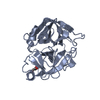
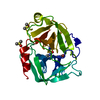
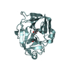
SHEET DETERMINATION METHOD: DSSP THE SHEETS PRESENTED AS "AA" IN EACH CHAIN ON SHEET RECORDS BELOW ... SHEET DETERMINATION METHOD: DSSP THE SHEETS PRESENTED AS "AA" IN EACH CHAIN ON SHEET RECORDS BELOW IS ACTUALLY AN 7-STRANDED BARREL THIS IS REPRESENTED BY A 8-STRANDED SHEET IN WHICH THE FIRST AND LAST STRANDS ARE IDENTICAL. THE SHEETS PRESENTED AS "AB" IN EACH CHAIN ON SHEET RECORDS BELOW IS ACTUALLY AN 6-STRANDED BARREL THIS IS REPRESENTED BY A 7-STRANDED SHEET IN WHICH THE FIRST AND LAST STRANDS ARE IDENTICAL.

-
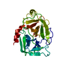
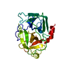
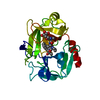
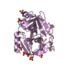
Structure visualization

Structure viewerMolecule:
MolmilJmol/JSmol

Downloads & links

-
Assembly

Deposited unit
A: COMPLEMENT FACTOR D
hetero molecules


Theoretical massNumber of molelcules
Total (without water)24,7915
Polymers24,4231
Non-polymers3684
Water5,423301
1


  • Idetical with deposited unit
  • defined by author&software
TypeNameSymmetry operationNumber
identity operation1_555x,y,z1
MethodPISA
Unit cell
Length a, b, c (Å)39.510, 49.550, 55.480
Angle α, β, γ (deg.)90.00, 105.64, 90.00
Int Tables number4
Space group name H-MP1211

-
Components

#1: Protein COMPLEMENT FACTOR D / ADIPSIN / C3 CONVERTASE ACTIVATOR / PROPERDIN FACTOR D


Mass: 24422.807 Da / Num. of mol.: 1 / Mutation: YES
Source method: isolated from a genetically manipulated source
Source: (gene. exp.) HOMO SAPIENS (human) / Plasmid: PUPE.05.05 / Cell line (production host): HEK293-E / Production host: HOMO SAPIENS (human) / References: UniProt: P00746, complement factor D
#2: Chemical
ChemComp-GOL / GLYCEROL / GLYCERIN / PROPANE-1,2,3-TRIOL


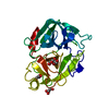
Mass: 92.094 Da / Num. of mol.: 4 / Source method: obtained synthetically / Formula: C3H8O3
#3: Water ChemComp-HOH / water


Mass: 18.015 Da / Num. of mol.: 301 / Source method: isolated from a natural source / Formula: H2O
Compound detailsENGINEERED RESIDUE IN CHAIN A, SER 208 TO ALA
Sequence detailsRESIDUE NUMBERING IN COORDINATES REFERS TO MATURE PROTEIN. MUTATION (SEE REMARK 400) IS SER183 TO ...RESIDUE NUMBERING IN COORDINATES REFERS TO MATURE PROTEIN. MUTATION (SEE REMARK 400) IS SER183 TO ALA IN COORDINATES.

-
Experimental details

-
Experiment

ExperimentMethod: X-RAY DIFFRACTION / Number of used crystals: 1

-
Sample preparation

CrystalDensity Matthews: 2.17 Å3/Da / Density % sol: 43.5 % / Description: NONE
Crystal growpH: 6 / Details: pH 6.0

-
Data collection

DiffractionMean temperature: 100 K
Diffraction sourceSource: SYNCHROTRON / Site: SLS / Beamline: X06SA / Wavelength: 1
DetectorType: DECTRIS PILATUS 6M / Detector: PIXEL / Date: Nov 30, 2009
RadiationProtocol: SINGLE WAVELENGTH / Monochromatic (M) / Laue (L): M / Scattering type: x-ray
Radiation wavelengthWavelength: 1 Å / Relative weight: 1
ReflectionResolution: 1.2→35.9 Å / Num. obs: 64058 / % possible obs: 99.3 % / Observed criterion σ(I): 3.5 / Redundancy: 2.9 % / Biso Wilson estimate: 8.535 Å2 / Rmerge(I) obs: 0.07 / Net I/σ(I): 8
Reflection shellResolution: 1.2→1.26 Å / Redundancy: 2.7 % / Rmerge(I) obs: 0.25 / Mean I/σ(I) obs: 3.5 / % possible all: 98.8

-
Processing

Software
NameVersionClassification
REFMAC5.5.0102refinement
iMOSFLMdata reduction
SCALAdata scaling
PHASERphasing
RefinementMethod to determine structure: MOLECULAR REPLACEMENT
Starting model: PDB ENTRY 1DSU
Resolution: 1.2→53.43 Å / Cor.coef. Fo:Fc: 0.972 / Cor.coef. Fo:Fc free: 0.959 / SU B: 1.038 / SU ML: 0.022 / Cross valid method: THROUGHOUT / ESU R: 0.04 / ESU R Free: 0.04 / Stereochemistry target values: MAXIMUM LIKELIHOOD / Details: HYDROGENS HAVE BEEN ADDED IN THE RIDING POSITIONS.
RfactorNum. reflection% reflectionSelection details
Rfree0.17605 3235 5.1 %RANDOM
Rwork0.14364 ---
obs0.14528 60768 99.22 %-
Solvent computationIon probe radii: 0.8 Å / Shrinkage radii: 0.8 Å / VDW probe radii: 1.4 Å / Solvent model: MASK
Displacement parametersBiso mean: 9.901 Å2
Baniso -1Baniso -2Baniso -3
1--0.16 Å20 Å2-0.06 Å2
2--0.22 Å20 Å2
3----0.09 Å2
Refinement stepCycle: LAST / Resolution: 1.2→53.43 Å
ProteinNucleic acidLigandSolventTotal
Num. atoms1711 0 24 301 2036
Refine LS restraints
Refine-IDTypeDev idealDev ideal targetNumber
X-RAY DIFFRACTIONr_bond_refined_d0.010.0211897
X-RAY DIFFRACTIONr_bond_other_d0.0010.021314
X-RAY DIFFRACTIONr_angle_refined_deg1.4561.982608
X-RAY DIFFRACTIONr_angle_other_deg0.93133221
X-RAY DIFFRACTIONr_dihedral_angle_1_deg5.8495267
X-RAY DIFFRACTIONr_dihedral_angle_2_deg34.11822.65879
X-RAY DIFFRACTIONr_dihedral_angle_3_deg13.73615313
X-RAY DIFFRACTIONr_dihedral_angle_4_deg18.261520
X-RAY DIFFRACTIONr_chiral_restr0.0920.2294
X-RAY DIFFRACTIONr_gen_planes_refined0.0070.0212157
X-RAY DIFFRACTIONr_gen_planes_other0.0010.02371
X-RAY DIFFRACTIONr_nbd_refined
X-RAY DIFFRACTIONr_nbd_other
X-RAY DIFFRACTIONr_nbtor_refined
X-RAY DIFFRACTIONr_nbtor_other
X-RAY DIFFRACTIONr_xyhbond_nbd_refined
X-RAY DIFFRACTIONr_xyhbond_nbd_other
X-RAY DIFFRACTIONr_metal_ion_refined
X-RAY DIFFRACTIONr_metal_ion_other
X-RAY DIFFRACTIONr_symmetry_vdw_refined
X-RAY DIFFRACTIONr_symmetry_vdw_other
X-RAY DIFFRACTIONr_symmetry_hbond_refined
X-RAY DIFFRACTIONr_symmetry_hbond_other
X-RAY DIFFRACTIONr_symmetry_metal_ion_refined
X-RAY DIFFRACTIONr_symmetry_metal_ion_other
X-RAY DIFFRACTIONr_mcbond_it1.2761.51198
X-RAY DIFFRACTIONr_mcbond_other0.3681.5485
X-RAY DIFFRACTIONr_mcangle_it2.1421943
X-RAY DIFFRACTIONr_mcangle_other
X-RAY DIFFRACTIONr_scbond_it2.8833699
X-RAY DIFFRACTIONr_scbond_other
X-RAY DIFFRACTIONr_scangle_it4.3474.5645
X-RAY DIFFRACTIONr_scangle_other
X-RAY DIFFRACTIONr_long_range_B_refined
X-RAY DIFFRACTIONr_long_range_B_other
X-RAY DIFFRACTIONr_rigid_bond_restr1.0933211
X-RAY DIFFRACTIONr_sphericity_free
X-RAY DIFFRACTIONr_sphericity_bonded
LS refinement shellResolution: 1.2→1.231 Å / Total num. of bins used: 20
RfactorNum. reflection% reflection
Rfree0.205 247 -
Rwork0.179 4418 -
obs--98.9 %
Refinement TLS params.Method: refined / Origin x: -1.219 Å / Origin y: 1.557 Å / Origin z: 13.138 Å
111213212223313233
T0.0275 Å2-0.0004 Å20.0088 Å2-0.0202 Å2-0.0002 Å2--0.0138 Å2
L0.0554 °2-0.0127 °20.0112 °2-0.0231 °2-0.017 °2--0.0573 °2
S-0.0001 Å °-0.0012 Å °-0.0037 Å °-0.0006 Å °0.001 Å °0.0007 Å °-0.0023 Å °-0.0001 Å °-0.0009 Å °

+
About Yorodumi

-
News

-
Feb 9, 2022. New format data for meta-information of EMDB entries

New format data for meta-information of EMDB entries

  • Version 3 of the EMDB header file is now the official format.
  • The previous official version 1.9 will be removed from the archive.

Related info.:EMDB header

External links:wwPDB to switch to version 3 of the EMDB data model

-
Aug 12, 2020. Covid-19 info

Covid-19 info

URL: https://pdbjlvh1.pdbj.org/emnavi/covid19.php

New page: Covid-19 featured information page in EM Navigator.

Related info.:Covid-19 info / Mar 5, 2020. Novel coronavirus structure data

+
Mar 5, 2020. Novel coronavirus structure data

Novel coronavirus structure data

Related info.:Yorodumi Speices / Aug 12, 2020. Covid-19 info

External links:COVID-19 featured content - PDBj / Molecule of the Month (242):Coronavirus Proteases

+
Jan 31, 2019. EMDB accession codes are about to change! (news from PDBe EMDB page)

EMDB accession codes are about to change! (news from PDBe EMDB page)

  • The allocation of 4 digits for EMDB accession codes will soon come to an end. Whilst these codes will remain in use, new EMDB accession codes will include an additional digit and will expand incrementally as the available range of codes is exhausted. The current 4-digit format prefixed with “EMD-” (i.e. EMD-XXXX) will advance to a 5-digit format (i.e. EMD-XXXXX), and so on. It is currently estimated that the 4-digit codes will be depleted around Spring 2019, at which point the 5-digit format will come into force.
  • The EM Navigator/Yorodumi systems omit the EMD- prefix.

Related info.:Q: What is EMD? / ID/Accession-code notation in Yorodumi/EM Navigator

External links:EMDB Accession Codes are Changing Soon! / Contact to PDBj

+
Jul 12, 2017. Major update of PDB

Major update of PDB

  • wwPDB released updated PDB data conforming to the new PDBx/mmCIF dictionary.
  • This is a major update changing the version number from 4 to 5, and with Remediation, in which all the entries are updated.
  • In this update, many items about electron microscopy experimental information are reorganized (e.g. em_software).
  • Now, EM Navigator and Yorodumi are based on the updated data.

External links:wwPDB Remediation / Enriched Model Files Conforming to OneDep Data Standards Now Available in the PDB FTP Archive

-
Yorodumi

Thousand views of thousand structures

  • Yorodumi is a browser for structure data from EMDB, PDB, SASBDB, etc.
  • This page is also the successor to EM Navigator detail page, and also detail information page/front-end page for Omokage search.
  • The word "yorodu" (or yorozu) is an old Japanese word meaning "ten thousand". "mi" (miru) is to see.

Related info.:EMDB / PDB / SASBDB / Comparison of 3 databanks / Yorodumi Search / Aug 31, 2016. New EM Navigator & Yorodumi / Yorodumi Papers / Jmol/JSmol / Function and homology information / Changes in new EM Navigator and Yorodumi

Read more