 Movie
Movie Controller


+ Open data

- Basic information
| Entry | Database: PDB / ID: 1hfd | ||||||
|---|---|---|---|---|---|---|---|
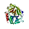
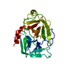
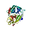
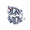
| Title | HUMAN COMPLEMENT FACTOR D IN A P21 CRYSTAL FORM | ||||||
 Components Components | COMPLEMENT FACTOR D | ||||||
 Keywords Keywords | SERINE PROTEASE / HYDROLASE / COMPLEMENT / FACTOR D / CATALYTIC TRIAD / SELF-REGULATION | ||||||
| Function / homology |  Function and homology information Function and homology informationcomplement factor D / Alternative complement activation / complement activation, alternative pathway / complement activation / zymogen activation / platelet alpha granule lumen / serine-type peptidase activity / response to bacterium / protein maturation / Platelet degranulation ...complement factor D / Alternative complement activation / complement activation, alternative pathway / complement activation / zymogen activation / platelet alpha granule lumen / serine-type peptidase activity / response to bacterium / protein maturation / Platelet degranulation / secretory granule lumen / ficolin-1-rich granule lumen / serine-type endopeptidase activity / Neutrophil degranulation / proteolysis / : / extracellular exosome / extracellular region Similarity search - Function | ||||||
| Biological species |  Homo sapiens (human) Homo sapiens (human) | ||||||
| Method |  X-RAY DIFFRACTION / MOLECULAR REPLACEMENT / Resolution: 2.3 Å X-RAY DIFFRACTION / MOLECULAR REPLACEMENT / Resolution: 2.3 Å | ||||||
 Authors Authors | Jing, H. / Babu, Y.S. / Moore, D. / Kilpatrick, J.M. / Liu, X.-Y. / Volanakis, J.E. / Narayana, S.V.L. | ||||||
 Citation Citation | Journal: J.Mol.Biol. / Year: 1998 Title: Structures of native and complexed complement factor D: implications of the atypical His57 conformation and self-inhibitory loop in the regulation of specific serine protease activity. Authors: Jing, H. / Babu, Y.S. / Moore, D. / Kilpatrick, J.M. / Liu, X.Y. / Volanakis, J.E. / Narayana, S.V. #1: Journal: Protein Sci. / Year: 1996Title: Complement Factor D, a Novel Serine Protease Authors: Volanakis, J.E. / Narayana, S.V. #2: Journal: J.Biol.Chem. / Year: 1995Title: Crystal Structure of a Complement Factor D Mutant Expressing Enhanced Catalytic Activity Authors: Kim, S. / Narayana, S.V. / Volanakis, J.E. #3: Journal: J.Mol.Biol. / Year: 1994Title: Structure of Human Factor D. A Complement System Protein at 2.0 A Resolution Authors: Narayana, S.V. / Carson, M. / El-Kabbani, O. / Kilpatrick, J.M. / Moore, D. / Chen, X. / Bugg, C.E. / Volanakis, J.E. / Delucas, L.J. #4: Journal: J.Mol.Biol. / Year: 1991Title: Crystallization and Preliminary X-Ray Investigation of Factor D of Human Complement Authors: Narayana, S.V. / Kilpatrick, J.M. / El-Kabbani, O. / Babu, Y.S. / Bugg, C.E. / Volanakis, J.E. / Delucas, L.J. | ||||||
| History |
|
- Structure visualization
| Structure viewer | Molecule: MolmilJmol/JSmol |
|---|
- Downloads & links
-Download
| PDBx/mmCIF format | 1hfd.cif.gz | 56.4 KB | Display | PDBx/mmCIF format |
|---|---|---|---|---|
| PDB format | pdb1hfd.ent.gz | 40.5 KB | Display | PDB format |
| PDBx/mmJSON format | 1hfd.json.gz | Tree view | PDBx/mmJSON format | |
| Others |  Other downloads Other downloads |
-Validation report
| Arichive directory | https://data.pdbj.org/pub/pdb/validation_reports/hf/1hfdftp://data.pdbj.org/pub/pdb/validation_reports/hf/1hfd | HTTPS FTP |
|---|
-Related structure data
| Related structure data |  1bioC  1dsuS S: Starting model for refinement C: citing same article ( |
|---|---|
| Similar structure data |


-Links
 PDBj
PDBj- Assembly
| Deposited unit | 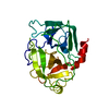
| ||||||||
|---|---|---|---|---|---|---|---|---|---|
| 1 |
| ||||||||
| Unit cell |
|
-Components
| #1: Protein | Mass: 24438.807 Da / Num. of mol.: 1 / Source method: isolated from a natural source / Source: (natural) Homo sapiens (human) / References: UniProt: P00746, complement factor D |
|---|---|
| #2: Water | ChemComp-HOH /  Mass: 18.015 Da / Num. of mol.: 75 / Source method: isolated from a natural source / Formula: H2O Mass: 18.015 Da / Num. of mol.: 75 / Source method: isolated from a natural source / Formula: H2O |
| Has protein modification | Y |
-Experimental details
-Experiment
| Experiment | Method: X-RAY DIFFRACTION / Number of used crystals: 4 |
|---|
- Sample preparation
| Crystal | Density Matthews: 2.15 Å3/Da / Density % sol: 44 % | ||||||||||||||||||||||||||||||||||||||||||
|---|---|---|---|---|---|---|---|---|---|---|---|---|---|---|---|---|---|---|---|---|---|---|---|---|---|---|---|---|---|---|---|---|---|---|---|---|---|---|---|---|---|---|---|
| Crystal grow | pH: 5.4 / Details: pH 5.4 | ||||||||||||||||||||||||||||||||||||||||||
| Crystal grow | *PLUS Method: vapor diffusion, hanging dropDetails: drop consists of equal volume of protein and reservoir solutions | ||||||||||||||||||||||||||||||||||||||||||
| Components of the solutions | *PLUS
|
-Data collection
| Diffraction | Mean temperature: 293 K |
|---|---|
| Diffraction source | Source: ROTATING ANODE / Type: RIGAKU RUH2R / Wavelength: 1.5418 |
| Detector | Type: SIEMENS / Detector: AREA DETECTOR / Date: Jun 1, 1993 / Details: COLLIMATOR |
| Radiation | Monochromator: GRAPHITE(002) / Monochromatic (M) / Laue (L): M / Scattering type: x-ray |
| Radiation wavelength | Wavelength: 1.5418 Å / Relative weight: 1 |
| Reflection | Resolution: 2.3→20 Å / Num. obs: 9267 / % possible obs: 95 % / Observed criterion σ(I): 2 / Redundancy: 4 % / Biso Wilson estimate: 15.7 Å2 / Rmerge(I) obs: 0.083 / Net I/σ(I): 15 |
| Reflection shell | Resolution: 2.3→2.4 Å / Redundancy: 4 % / Rmerge(I) obs: 0.242 / Mean I/σ(I) obs: 2.8 / % possible all: 79 |
| Reflection | *PLUS Num. measured all: 36098 |
| Reflection shell | *PLUS % possible obs: 79 % |
- Processing
| Software |
| ||||||||||||||||||||||||||||||||||||||||||||||||||||||||||||
|---|---|---|---|---|---|---|---|---|---|---|---|---|---|---|---|---|---|---|---|---|---|---|---|---|---|---|---|---|---|---|---|---|---|---|---|---|---|---|---|---|---|---|---|---|---|---|---|---|---|---|---|---|---|---|---|---|---|---|---|---|---|
| Refinement | Method to determine structure: MOLECULAR REPLACEMENT Starting model: PDB ENTRY 1DSU MOLECULE B Resolution: 2.3→20 Å / Rfactor Rfree error: 0.007 / Data cutoff high absF: 1000000 / Data cutoff low absF: 0.001 / Cross valid method: THROUGHOUT / σ(F): 2 / Details: BULK SOLVENT CORRECTION WAS APPLIED
| ||||||||||||||||||||||||||||||||||||||||||||||||||||||||||||
| Displacement parameters | Biso mean: 22 Å2 | ||||||||||||||||||||||||||||||||||||||||||||||||||||||||||||
| Refine analyze |
| ||||||||||||||||||||||||||||||||||||||||||||||||||||||||||||
| Refinement step | Cycle: LAST / Resolution: 2.3→20 Å
| ||||||||||||||||||||||||||||||||||||||||||||||||||||||||||||
| Refine LS restraints |
| ||||||||||||||||||||||||||||||||||||||||||||||||||||||||||||
| LS refinement shell | Resolution: 2.3→2.4 Å / Rfactor Rfree error: 0.027 / Total num. of bins used: 8
| ||||||||||||||||||||||||||||||||||||||||||||||||||||||||||||
| Xplor file |
| ||||||||||||||||||||||||||||||||||||||||||||||||||||||||||||
| Software | *PLUS Name: X-PLOR / Version: 3.851 / Classification: refinement | ||||||||||||||||||||||||||||||||||||||||||||||||||||||||||||
| Refinement | *PLUS | ||||||||||||||||||||||||||||||||||||||||||||||||||||||||||||
| Solvent computation | *PLUS | ||||||||||||||||||||||||||||||||||||||||||||||||||||||||||||
| Displacement parameters | *PLUS | ||||||||||||||||||||||||||||||||||||||||||||||||||||||||||||
| Refine LS restraints | *PLUS
|
