ムービー
ムービー コントローラー


+ データを開く

- 基本情報
基本情報
| 登録情報 | データベース: PDB / ID: 2xvr | ||||||
|---|---|---|---|---|---|---|---|
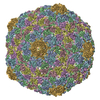
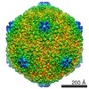
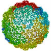
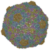
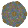
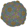
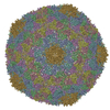
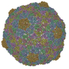
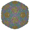
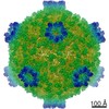
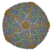
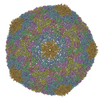
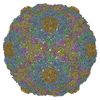
| タイトル | Phage T7 empty mature head shell | ||||||
 要素 要素 | MAJOR CAPSID PROTEIN 10A | ||||||
 キーワード キーワード | VIRUS / CAPSID MATURATION / MORPHOGENETIC INTERMEDIATE | ||||||
| 機能・相同性 | Capsid Gp10A/Gp10B / : / Major capsid protein / viral capsid / viral translational frameshifting / identical protein binding / Major capsid protein 機能・相同性情報 機能・相同性情報 | ||||||
| 生物種 |   ENTEROBACTERIA PHAGE T7 (ファージ) ENTEROBACTERIA PHAGE T7 (ファージ) | ||||||
| 手法 | 電子顕微鏡法 / 単粒子再構成法 / クライオ電子顕微鏡法 / 解像度: 10.8 Å | ||||||
 データ登録者 データ登録者 | Ionel, A. / Velazquez-Muriel, J.A. / Luque, D. / Cuervo, A. / Caston, J.R. / Valpuesta, J.M. / Martin-Benito, J. / Carrascosa, J.L. | ||||||
 引用 引用 | ジャーナル: J Biol Chem / 年: 2011 タイトル: Molecular rearrangements involved in the capsid shell maturation of bacteriophage T7. 著者: Alina Ionel / Javier A Velázquez-Muriel / Daniel Luque / Ana Cuervo / José R Castón / José M Valpuesta / Jaime Martín-Benito / José L Carrascosa /  要旨: Maturation of dsDNA bacteriophages involves assembling the virus prohead from a limited set of structural components followed by rearrangements required for the stability that is necessary for ...Maturation of dsDNA bacteriophages involves assembling the virus prohead from a limited set of structural components followed by rearrangements required for the stability that is necessary for infecting a host under challenging environmental conditions. Here, we determine the mature capsid structure of T7 at 1 nm resolution by cryo-electron microscopy and compare it with the prohead to reveal the molecular basis of T7 shell maturation. The mature capsid presents an expanded and thinner shell, with a drastic rearrangement of the major protein monomers that increases in their interacting surfaces, in turn resulting in a new bonding lattice. The rearrangements include tilting, in-plane rotation, and radial expansion of the subunits, as well as a relative bending of the A- and P-domains of each subunit. The unique features of this shell transformation, which does not employ the accessory proteins, inserted domains, or molecular interactions observed in other phages, suggest a simple capsid assembling strategy that may have appeared early in the evolution of these viruses. | ||||||
| 履歴 |
|
- 構造の表示
構造の表示
| ムービー |
ムービービューア |
|---|---|
| 構造ビューア | 分子: MolmilJmol/JSmol |
- ダウンロードとリンク
-ダウンロード
| PDBx/mmCIF形式 | 2xvr.cif.gz | 288.3 KB | 表示 | PDBx/mmCIF形式 |
|---|---|---|---|---|
| PDB形式 | pdb2xvr.ent.gz | 225.4 KB | 表示 | PDB形式 |
| PDBx/mmJSON形式 | 2xvr.json.gz | ツリー表示 | PDBx/mmJSON形式 | |
| その他 |  その他のダウンロード その他のダウンロード |
-検証レポート
| アーカイブディレクトリ | https://data.pdbj.org/pub/pdb/validation_reports/xv/2xvrftp://data.pdbj.org/pub/pdb/validation_reports/xv/2xvr | HTTPS FTP |
|---|
-関連構造データ
-リンク
 PDBj
PDBj- 集合体
集合体
| 登録構造単位 | 
|
|---|---|
| 1 | x 60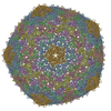
|
| 2 |
|
| 3 | x 5
|
| 4 | x 6
|
| 5 | 
|
| 対称性 | 点対称性: (シェーンフリース記号: I (正20面体型対称)) |
-要素
| #1: タンパク質 | 分子量: 36589.625 Da / 分子数: 7 / 由来タイプ: 天然 / 由来: (天然) ENTEROBACTERIA PHAGE T7 (ファージ) / 参照: UniProt: P19726 |
|---|
-実験情報
-実験
| 実験 | 手法: 電子顕微鏡法 |
|---|---|
| EM実験 | 試料の集合状態: PARTICLE / 3次元再構成法: 単粒子再構成法 |
- 試料調製
試料調製
| 構成要素 | 名称: PHAGE T7 EMPTY HEAD / タイプ: VIRUS |
|---|---|
| 緩衝液 | 名称: 50 MM TRIS-HCL, PH 7.8, 10 MM MGCL2, 0.1 M NACL / pH: 7.8 / 詳細: 50 MM TRIS-HCL, PH 7.8, 10 MM MGCL2, 0.1 M NACL |
| 試料 | 包埋: NO / シャドウイング: NO / 染色: NO / 凍結: YES |
| 試料支持 | 詳細: HOLEY CARBON |
| 急速凍結 | 凍結剤: ETHANE / 詳細: VITRIFICATION 1 -- CRYOGEN- ETHANE, |
- 電子顕微鏡撮影
| 実験機器 |  モデル: Tecnai F20 / 画像提供: FEI Company |
|---|---|
| 顕微鏡 | モデル: FEI TECNAI F20 |
| 電子銃 | 電子線源:  FIELD EMISSION GUN / 加速電圧: 200 kV / 照射モード: FLOOD BEAM FIELD EMISSION GUN / 加速電圧: 200 kV / 照射モード: FLOOD BEAM |
| 電子レンズ | モード: BRIGHT FIELD / 倍率(公称値): 50000 X |
| 撮影 | フィルム・検出器のモデル: KODAK SO-163 FILM |
- 解析
解析
| EMソフトウェア | 名称: Xmipp / カテゴリ: 3次元再構成 | ||||||||||||
|---|---|---|---|---|---|---|---|---|---|---|---|---|---|
| 対称性 | 点対称性: I (正20面体型対称) | ||||||||||||
| 3次元再構成 | 解像度: 10.8 Å / 粒子像の数: 5100 / ピクセルサイズ(実測値): 1.4 Å 詳細: SUBMISSION BASED ON EXPERIMENTAL DATA FROM EMDB EMD-1810. (DEPOSITION ID: 7638). 対称性のタイプ: POINT | ||||||||||||
| 原子モデル構築 | 空間: REAL | ||||||||||||
| 原子モデル構築 | PDB-ID: 1OHG Accession code: 1OHG / Source name: PDB / タイプ: experimental model | ||||||||||||
| 精密化 | 最高解像度: 10.8 Å | ||||||||||||
| 精密化ステップ | サイクル: LAST / 最高解像度: 10.8 Å
|