 Movie
Movie Controller


+ Open data

- Basic information
| Entry | Database: PDB / ID: 2xj4 | ||||||
|---|---|---|---|---|---|---|---|
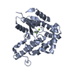
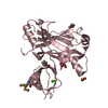
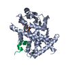
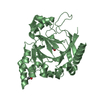
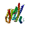
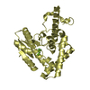
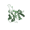
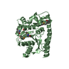
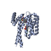
| Title | Structure of the bacterial cell division regulator protein MipZ | ||||||
 Components Components | MIPZ | ||||||
 Keywords Keywords | REPLICATION / CELL DIVISION / ATPASE / WACA | ||||||
| Function / homology |  Function and homology information Function and homology informationATPase MipZ / ATPase MipZ / : / P-loop containing nucleotide triphosphate hydrolases / Rossmann fold / P-loop containing nucleoside triphosphate hydrolase / 3-Layer(aba) Sandwich / Alpha Beta Similarity search - Domain/homology | ||||||
| Biological species |  CAULOBACTER CRESCENTUS (bacteria) CAULOBACTER CRESCENTUS (bacteria) | ||||||
| Method |  X-RAY DIFFRACTION / SYNCHROTRON / SAD / Resolution: 1.6 Å X-RAY DIFFRACTION / SYNCHROTRON / SAD / Resolution: 1.6 Å | ||||||
 Authors Authors | Michie, K.A. / Lowe, J. | ||||||
 Citation Citation | Journal: Mol.Cell / Year: 2012 Title: Localized Dimerization and Nucleoid Binding Drive Gradient Formation by the Bacterial Cell Division Inhibitor Mipz. Authors: Kiekebusch, D. / Michie, K.A. / Essen, L.O. / Lowe, J. / Thanbichler, M. | ||||||
| History |
|
- Structure visualization
| Structure viewer | Molecule: MolmilJmol/JSmol |
|---|
- Downloads & links
-Download
| PDBx/mmCIF format | 2xj4.cif.gz | 132 KB | Display | PDBx/mmCIF format |
|---|---|---|---|---|
| PDB format | pdb2xj4.ent.gz | 104.4 KB | Display | PDB format |
| PDBx/mmJSON format | 2xj4.json.gz | Tree view | PDBx/mmJSON format | |
| Others |  Other downloads Other downloads |
-Validation report
| Arichive directory | https://data.pdbj.org/pub/pdb/validation_reports/xj/2xj4ftp://data.pdbj.org/pub/pdb/validation_reports/xj/2xj4 | HTTPS FTP |
|---|
-Related structure data


-Links
 PDBj
PDBj- Assembly
| Deposited unit | 
| ||||||||
|---|---|---|---|---|---|---|---|---|---|
| 1 |
| ||||||||
| Unit cell |
| ||||||||
| Components on special symmetry positions |
|
-Components
| #1: Protein | Mass: 31763.359 Da / Num. of mol.: 1 Source method: isolated from a genetically manipulated source Source: (gene. exp.) CAULOBACTER CRESCENTUS (bacteria) / Strain: CB15N / Description: PCR OF GENOMIC DNA SUPPLIED BY ATCC (19089D) / Plasmid: PHIS17 / Production host: |
|---|---|
| #2: Water | ChemComp-HOH /  Mass: 18.015 Da / Num. of mol.: 353 / Source method: isolated from a natural source / Formula: H2O Mass: 18.015 Da / Num. of mol.: 353 / Source method: isolated from a natural source / Formula: H2O |
-Experimental details
-Experiment
| Experiment | Method: X-RAY DIFFRACTION / Number of used crystals: 1 |
|---|
- Sample preparation
| Crystal | Density Matthews: 2.5 Å3/Da / Density % sol: 50.7 % / Description: NONE |
|---|---|
| Crystal grow | pH: 6.5 / Details: 0.1 M MES/NAOH PH6.5, 30% V/V PEG400 |
-Data collection
| Diffraction | Mean temperature: 100 K |
|---|---|
| Diffraction source | Source: SYNCHROTRON / Site: ESRF  / Beamline: ID23-1 / Wavelength: 0.9747 / Beamline: ID23-1 / Wavelength: 0.9747 |
| Detector | Type: ADSC QUANTUM 315r / Detector: CCD / Date: Jun 16, 2007 |
| Radiation | Protocol: SINGLE WAVELENGTH / Monochromatic (M) / Laue (L): M / Scattering type: x-ray |
| Radiation wavelength | Wavelength: 0.9747 Å / Relative weight: 1 |
| Reflection | Resolution: 1.5→20.69 Å / Num. obs: 40200 / % possible obs: 99.6 % / Observed criterion σ(I): 0 / Redundancy: 5.4 % / Biso Wilson estimate: 18.813 Å2 / Rmerge(I) obs: 0.09 / Net I/σ(I): 11.6 |
| Reflection shell | Resolution: 1.6→1.69 Å / Redundancy: 5.5 % / Rmerge(I) obs: 0.29 / Mean I/σ(I) obs: 5.1 / % possible all: 100 |
- Processing
| Software |
| ||||||||||||||||||||||||||||||||||||||||||||||||||||||||||||||||||||||||||||||||||||||||||||||||||||||||||||||||||||||||||||||||||||||||||||||||||||||||||||||||||||||||||||||||||||||
|---|---|---|---|---|---|---|---|---|---|---|---|---|---|---|---|---|---|---|---|---|---|---|---|---|---|---|---|---|---|---|---|---|---|---|---|---|---|---|---|---|---|---|---|---|---|---|---|---|---|---|---|---|---|---|---|---|---|---|---|---|---|---|---|---|---|---|---|---|---|---|---|---|---|---|---|---|---|---|---|---|---|---|---|---|---|---|---|---|---|---|---|---|---|---|---|---|---|---|---|---|---|---|---|---|---|---|---|---|---|---|---|---|---|---|---|---|---|---|---|---|---|---|---|---|---|---|---|---|---|---|---|---|---|---|---|---|---|---|---|---|---|---|---|---|---|---|---|---|---|---|---|---|---|---|---|---|---|---|---|---|---|---|---|---|---|---|---|---|---|---|---|---|---|---|---|---|---|---|---|---|---|---|---|
| Refinement | Method to determine structure: SAD Starting model: NONE Resolution: 1.6→61.31 Å / Cor.coef. Fo:Fc: 0.976 / Cor.coef. Fo:Fc free: 0.962 / SU B: 2.612 / SU ML: 0.042 / Cross valid method: THROUGHOUT / ESU R: 0.08 / ESU R Free: 0.072 / Stereochemistry target values: MAXIMUM LIKELIHOOD / Details: HYDROGENS HAVE BEEN ADDED IN THE RIDING POSITIONS.
| ||||||||||||||||||||||||||||||||||||||||||||||||||||||||||||||||||||||||||||||||||||||||||||||||||||||||||||||||||||||||||||||||||||||||||||||||||||||||||||||||||||||||||||||||||||||
| Solvent computation | Ion probe radii: 0.8 Å / Shrinkage radii: 0.8 Å / VDW probe radii: 1.4 Å / Solvent model: BABINET MODEL WITH MASK | ||||||||||||||||||||||||||||||||||||||||||||||||||||||||||||||||||||||||||||||||||||||||||||||||||||||||||||||||||||||||||||||||||||||||||||||||||||||||||||||||||||||||||||||||||||||
| Displacement parameters | Biso mean: 23.685 Å2
| ||||||||||||||||||||||||||||||||||||||||||||||||||||||||||||||||||||||||||||||||||||||||||||||||||||||||||||||||||||||||||||||||||||||||||||||||||||||||||||||||||||||||||||||||||||||
| Refinement step | Cycle: LAST / Resolution: 1.6→61.31 Å
| ||||||||||||||||||||||||||||||||||||||||||||||||||||||||||||||||||||||||||||||||||||||||||||||||||||||||||||||||||||||||||||||||||||||||||||||||||||||||||||||||||||||||||||||||||||||
| Refine LS restraints |
|