 Movie
Movie Controller


+ Open data

- Basic information
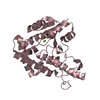
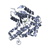
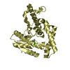
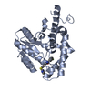
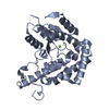
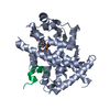
| Entry | Database: PDB / ID: 3hb0 | ||||||
|---|---|---|---|---|---|---|---|
| Title | Structure of edeya2 complexed with bef3 | ||||||
 Components Components | Eyes absent homolog 2 (Drosophila) | ||||||
 Keywords Keywords | HYDROLASE / alpha/beta hydrolase | ||||||
| Function / homology |  Function and homology information Function and homology informationmesodermal cell fate specification / striated muscle tissue development / histone H2AXY142 phosphatase activity / mitochondrial outer membrane permeabilization / negative regulation of extrinsic apoptotic signaling pathway in absence of ligand / extrinsic apoptotic signaling pathway in absence of ligand / protein-tyrosine-phosphatase / protein tyrosine phosphatase activity / positive regulation of DNA repair / Recruitment and ATM-mediated phosphorylation of repair and signaling proteins at DNA double strand breaks ...mesodermal cell fate specification / striated muscle tissue development / histone H2AXY142 phosphatase activity / mitochondrial outer membrane permeabilization / negative regulation of extrinsic apoptotic signaling pathway in absence of ligand / extrinsic apoptotic signaling pathway in absence of ligand / protein-tyrosine-phosphatase / protein tyrosine phosphatase activity / positive regulation of DNA repair / Recruitment and ATM-mediated phosphorylation of repair and signaling proteins at DNA double strand breaks / cell differentiation / DNA repair / magnesium ion binding / DNA-templated transcription / mitochondrion / nucleoplasm / nucleus / cytosol Similarity search - Function | ||||||
| Biological species |  Homo sapiens (human) Homo sapiens (human) | ||||||
| Method |  X-RAY DIFFRACTION / SYNCHROTRON / MOLECULAR REPLACEMENT / Resolution: 2.5 Å X-RAY DIFFRACTION / SYNCHROTRON / MOLECULAR REPLACEMENT / Resolution: 2.5 Å | ||||||
 Authors Authors | Jung, S.K. / Jeong, D.G. / Ryu, S.E. / Kim, S.J. | ||||||
 Citation Citation | Journal: Faseb J. / Year: 2010 Title: Crystal structure of ED-Eya2: insight into dual roles as a protein tyrosine phosphatase and a transcription factor Authors: Jung, S.K. / Jeong, D.G. / Chung, S.J. / Kim, J.H. / Park, B.C. / Tonks, N.K. / Ryu, S.E. / Kim, S.J. | ||||||
| History |
|
- Structure visualization
| Structure viewer | Molecule: MolmilJmol/JSmol |
|---|
- Downloads & links
-Download
| PDBx/mmCIF format | 3hb0.cif.gz | 213.1 KB | Display | PDBx/mmCIF format |
|---|---|---|---|---|
| PDB format | pdb3hb0.ent.gz | 171.5 KB | Display | PDB format |
| PDBx/mmJSON format | 3hb0.json.gz | Tree view | PDBx/mmJSON format | |
| Others |  Other downloads Other downloads |
-Validation report
| Arichive directory | https://data.pdbj.org/pub/pdb/validation_reports/hb/3hb0ftp://data.pdbj.org/pub/pdb/validation_reports/hb/3hb0 | HTTPS FTP |
|---|
-Related structure data
| Related structure data |  3hb1C  3gebS C: citing same article ( S: Starting model for refinement |
|---|---|
| Similar structure data |


-Links
 PDBj
PDBj


- Assembly
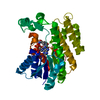
| Deposited unit | 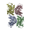
| ||||||||
|---|---|---|---|---|---|---|---|---|---|
| 1 | 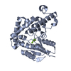
| ||||||||
| 2 | 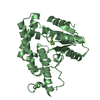
| ||||||||
| 3 | 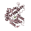
| ||||||||
| 4 | 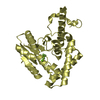
| ||||||||
| Unit cell |
|
-Components
| #1: Protein | Mass: 31068.354 Da / Num. of mol.: 4 / Fragment: Eya domain Source method: isolated from a genetically manipulated source Source: (gene. exp.) Homo sapiens (human) / Production host:  References: UniProt: Q86U84, UniProt: O00167*PLUS, protein-tyrosine-phosphatase #2: Chemical | ChemComp-BEF /   Mass: 66.007 Da / Num. of mol.: 4 / Source method: obtained synthetically / Formula: BeF3 Mass: 66.007 Da / Num. of mol.: 4 / Source method: obtained synthetically / Formula: BeF3#3: Chemical | ChemComp-MG /   Mass: 24.305 Da / Num. of mol.: 4 / Source method: obtained synthetically / Formula: Mg Mass: 24.305 Da / Num. of mol.: 4 / Source method: obtained synthetically / Formula: Mg#4: Water | ChemComp-HOH / |  Mass: 18.015 Da / Num. of mol.: 139 / Source method: isolated from a natural source / Formula: H2O Mass: 18.015 Da / Num. of mol.: 139 / Source method: isolated from a natural source / Formula: H2O |
|---|
-Experimental details
-Experiment
| Experiment | Method: X-RAY DIFFRACTION / Number of used crystals: 1 |
|---|
- Sample preparation
| Crystal | Density Matthews: 4.08 Å3/Da / Density % sol: 69.89 % |
|---|---|
| Crystal grow | Temperature: 291 K / Method: vapor diffusion, sitting drop / pH: 6 Details: 2.4M NaCl, pH 6.0, VAPOR DIFFUSION, SITTING DROP, temperature 291K |
-Data collection
| Diffraction | Mean temperature: 100 K |
|---|---|
| Diffraction source | Source: SYNCHROTRON / Site: SPring-8  / Beamline: BL38B1 / Wavelength: 1 Å / Beamline: BL38B1 / Wavelength: 1 Å |
| Detector | Detector: CCD |
| Radiation | Protocol: SINGLE WAVELENGTH / Monochromatic (M) / Laue (L): M / Scattering type: x-ray |
| Radiation wavelength | Wavelength: 1 Å / Relative weight: 1 |
| Reflection | Resolution: 2.5→40 Å / Num. all: 69154 / Num. obs: 68947 / % possible obs: 99.7 % / Observed criterion σ(F): 0 / Observed criterion σ(I): 0 / Rmerge(I) obs: 0.06 / Rsym value: 0.074 / Net I/σ(I): 26.9 |
| Reflection shell | Resolution: 2.5→2.59 Å / Mean I/σ(I) obs: 2.4 / Num. unique all: 6733 / Num. unique obs: 6842 / Rsym value: 0.386 |
- Processing
| Software |
| |||||||||||||||||||||||||
|---|---|---|---|---|---|---|---|---|---|---|---|---|---|---|---|---|---|---|---|---|---|---|---|---|---|---|
| Refinement | Method to determine structure: MOLECULAR REPLACEMENT Starting model: PDB entry 3GEB Resolution: 2.5→40 Å / Isotropic thermal model: overall / Cross valid method: THROUGHOUT / σ(F): 0 / σ(I): 0 / Stereochemistry target values: Engh & Huber
| |||||||||||||||||||||||||
| Refinement step | Cycle: LAST / Resolution: 2.5→40 Å
|
