 Movie
Movie Controller

[English] 日本語
 Yorodumi
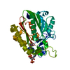
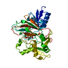
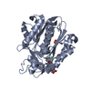
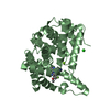
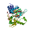
- PDB-3pg5: Crystal structure of protein DIP2308 from Corynebacterium diphthe... -
+ Open data

- Basic information
| Entry | Database: PDB / ID: 3pg5 | ||||||
|---|---|---|---|---|---|---|---|
| Title | Crystal structure of protein DIP2308 from Corynebacterium diphtheriae, Northeast Structural Genomics Consortium Target CdR78 | ||||||
 Components Components | Uncharacterized protein | ||||||
 Keywords Keywords | Structural genomics / Unknown function / PSI-Biology / Protein Structure Initiative / Northeast Structural Genomics Consortium / NESG / alpha-beta p-loop containing protein / ATP-binding protein | ||||||
| Function / homology | : / AAA domain / AAA domain / P-loop containing nucleotide triphosphate hydrolases / Rossmann fold / P-loop containing nucleoside triphosphate hydrolase / 3-Layer(aba) Sandwich / Alpha Beta / AAA domain-containing protein Function and homology information Function and homology information | ||||||
| Biological species |  Corynebacterium diphtheriae (bacteria) Corynebacterium diphtheriae (bacteria) | ||||||
| Method |  X-RAY DIFFRACTION / SYNCHROTRON / SAD / Resolution: 3.3 Å X-RAY DIFFRACTION / SYNCHROTRON / SAD / Resolution: 3.3 Å | ||||||
 Authors Authors | Forouhar, F. / Lew, S. / Seetharaman, J. / Lee, D. / Ciccosanti, C. / Sahdev, S. / Nair, R. / Rost, B. / Acton, T.B. / Xiao, R. ...Forouhar, F. / Lew, S. / Seetharaman, J. / Lee, D. / Ciccosanti, C. / Sahdev, S. / Nair, R. / Rost, B. / Acton, T.B. / Xiao, R. / Everett, J.K. / Montelione, G.T. / Hunt, J.F. / Tong, L. / Northeast Structural Genomics Consortium (NESG) | ||||||
 Citation Citation | Journal: To be Published Title: Crystal structure of protein DIP2308 from Corynebacterium diphtheriae, Northeast Structural Genomics Consortium Target CdR78 Authors: Forouhar, F. / Lew, S. / Seetharaman, J. / Lee, D. / Ciccosanti, C. / Sahdev, S. / Nair, R. / Rost, B. / Acton, T.B. / Xiao, R. / Everett, J.K. / Montelione, G.T. / Hunt, J.F. / Tong, L. | ||||||
| History |
|
- Structure visualization
| Structure viewer | Molecule: MolmilJmol/JSmol |
|---|
- Downloads & links
-Download
| PDBx/mmCIF format | 3pg5.cif.gz | 244.7 KB | Display | PDBx/mmCIF format |
|---|---|---|---|---|
| PDB format | pdb3pg5.ent.gz | 199.6 KB | Display | PDB format |
| PDBx/mmJSON format | 3pg5.json.gz | Tree view | PDBx/mmJSON format | |
| Others |  Other downloads Other downloads |
-Validation report
| Arichive directory | https://data.pdbj.org/pub/pdb/validation_reports/pg/3pg5ftp://data.pdbj.org/pub/pdb/validation_reports/pg/3pg5 | HTTPS FTP |
|---|
-Related structure data
| Similar structure data | |
|---|---|
| Other databases |
|


-Links
 PDBj
PDBj- Assembly
| Deposited unit | 
| ||||||||
|---|---|---|---|---|---|---|---|---|---|
| 1 | 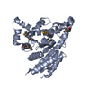
| ||||||||
| 2 | 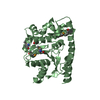
| ||||||||
| 3 | 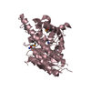
| ||||||||
| 4 | 
| ||||||||
| 5 | 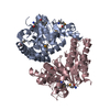
| ||||||||
| 6 | 
| ||||||||
| Unit cell |
| ||||||||
| Details | DIMER IN THE CRYSTAL, WHEREAS LIGHT SCATTERING DATA SHOWS MONOMERIC PROTEIN IN SOLUTION. |
-Components
| #1: Protein | Mass: 41062.555 Da / Num. of mol.: 4 Source method: isolated from a genetically manipulated source Source: (gene. exp.) Corynebacterium diphtheriae (bacteria) / Strain: NCTC 13129 / Gene: DIP2308 / Production host: #2: Water | ChemComp-HOH / |  Mass: 18.015 Da / Num. of mol.: 21 / Source method: isolated from a natural source / Formula: H2O Mass: 18.015 Da / Num. of mol.: 21 / Source method: isolated from a natural source / Formula: H2OHas protein modification | Y | |
|---|
-Experimental details
-Experiment
| Experiment | Method: X-RAY DIFFRACTION / Number of used crystals: 1 |
|---|
- Sample preparation
| Crystal | Density Matthews: 3.24 Å3/Da / Density % sol: 62.07 % |
|---|---|
| Crystal grow | Temperature: 277 K / Method: micro batch under oil / pH: 9 Details: Protein solution: 10 mM Tris (pH 7.5), 100 mM sodium chloride, and 5 mM DTT. Reservoir solution: 0.1 M TAPS (pH 9), 40% PEG400, and 0.1 M magnesium nitrate, micro batch under oil, temperature 277K |
-Data collection
| Diffraction | Mean temperature: 100 K |
|---|---|
| Diffraction source | Source: SYNCHROTRON / Site: NSLS  / Beamline: X4C / Wavelength: 0.97908 Å / Beamline: X4C / Wavelength: 0.97908 Å |
| Detector | Type: MAR CCD 165 mm / Detector: CCD / Date: Oct 12, 2010 / Details: mirrors |
| Radiation | Monochromator: Si 111 CHANNEL / Protocol: SINGLE WAVELENGTH / Monochromatic (M) / Laue (L): M / Scattering type: x-ray |
| Radiation wavelength | Wavelength: 0.97908 Å / Relative weight: 1 |
| Reflection | Resolution: 3.3→30 Å / Num. all: 62028 / Num. obs: 62028 / % possible obs: 100 % / Observed criterion σ(F): 0 / Observed criterion σ(I): 0 / Redundancy: 13.6 % / Biso Wilson estimate: 55.2 Å2 / Rmerge(I) obs: 0.23 / Rsym value: 0.187 / Net I/σ(I): 13.47 |
| Reflection shell | Resolution: 3.3→3.42 Å / Redundancy: 11 % / Rmerge(I) obs: 0.814 / Mean I/σ(I) obs: 2.56 / Num. unique all: 6231 / Rsym value: 0.738 / % possible all: 100 |
- Processing
| Software |
| |||||||||||||||||||||||||
|---|---|---|---|---|---|---|---|---|---|---|---|---|---|---|---|---|---|---|---|---|---|---|---|---|---|---|
| Refinement | Method to determine structure: SAD / Resolution: 3.3→20 Å / Rfactor Rfree error: 0.003 / Data cutoff high absF: 166084.6 / Data cutoff low absF: 0 / Isotropic thermal model: RESTRAINED / Cross valid method: THROUGHOUT / σ(F): 1 / σ(I): 1 / Stereochemistry target values: Engh & Huber
| |||||||||||||||||||||||||
| Solvent computation | Solvent model: FLAT MODEL / Bsol: 13.3223 Å2 / ksol: 0.3 e/Å3 | |||||||||||||||||||||||||
| Displacement parameters | Biso mean: 56.6 Å2
| |||||||||||||||||||||||||
| Refine analyze |
| |||||||||||||||||||||||||
| Refinement step | Cycle: LAST / Resolution: 3.3→20 Å
| |||||||||||||||||||||||||
| Refine LS restraints |
| |||||||||||||||||||||||||
| Refine LS restraints NCS | NCS model details: NONE | |||||||||||||||||||||||||
| LS refinement shell | Resolution: 3.3→3.42 Å / Rfactor Rfree error: 0.014 / Total num. of bins used: 10
|
