 ムービー
ムービー コントローラー


+ データを開く

- 基本情報
基本情報
| 登録情報 | データベース: PDB / ID: 2xj9 | ||||||
|---|---|---|---|---|---|---|---|
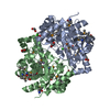
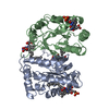
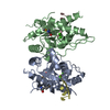
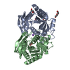
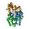
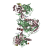
| タイトル | Dimer Structure of the bacterial cell division regulator MipZ | ||||||
 要素 要素 | MIPZ | ||||||
 キーワード キーワード | REPLICATION / CELL DIVISION / ATPASE / WACA | ||||||
| 機能・相同性 |  機能・相同性情報 機能・相同性情報 | ||||||
| 生物種 |  CAULOBACTER CRESCENTUS (バクテリア) CAULOBACTER CRESCENTUS (バクテリア) | ||||||
| 手法 |  X線回折 / シンクロトロン / 分子置換 / 解像度: 2.8 Å X線回折 / シンクロトロン / 分子置換 / 解像度: 2.8 Å | ||||||
 データ登録者 データ登録者 | Michie, K.A. / Lowe, J. | ||||||
 引用 引用 | ジャーナル: Mol.Cell / 年: 2012 タイトル: Localized Dimerization and Nucleoid Binding Drive Gradient Formation by the Bacterial Cell Division Inhibitor Mipz. 著者: Kiekebusch, D. / Michie, K.A. / Essen, L.O. / Lowe, J. / Thanbichler, M. | ||||||
| 履歴 |
|
- 構造の表示
構造の表示
| 構造ビューア | 分子: MolmilJmol/JSmol |
|---|
- ダウンロードとリンク
-ダウンロード
| PDBx/mmCIF形式 | 2xj9.cif.gz | 120.1 KB | 表示 | PDBx/mmCIF形式 |
|---|---|---|---|---|
| PDB形式 | pdb2xj9.ent.gz | 92.3 KB | 表示 | PDB形式 |
| PDBx/mmJSON形式 | 2xj9.json.gz | ツリー表示 | PDBx/mmJSON形式 | |
| その他 |  その他のダウンロード その他のダウンロード |
-検証レポート
| アーカイブディレクトリ | https://data.pdbj.org/pub/pdb/validation_reports/xj/2xj9ftp://data.pdbj.org/pub/pdb/validation_reports/xj/2xj9 | HTTPS FTP |
|---|
-関連構造データ


-リンク
 PDBj
PDBj
- 集合体
集合体
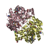
| 登録構造単位 | 
| ||||||||||||||||||
|---|---|---|---|---|---|---|---|---|---|---|---|---|---|---|---|---|---|---|---|
| 1 |
| ||||||||||||||||||
| 単位格子 |
| ||||||||||||||||||
| 非結晶学的対称性 (NCS) | NCSドメイン:
NCSドメイン領域:
NCS oper: (Code: given / Matrix: (1), |
-要素
| #1: タンパク質 | 分子量: 31719.348 Da / 分子数: 2 / 変異: YES / 由来タイプ: 組換発現 由来: (組換発現) CAULOBACTER CRESCENTUS (バクテリア)株: CB15N 解説: PCRED FROM GENOMIC DNA FROM ATCC 19089D. MUTATION CONSTRUCTED BY SPLICE-OVERLAP EXTENSION. プラスミド: PHIS17 / 発現宿主: #2: 化合物 |   分子量: 24.305 Da / 分子数: 2 / 由来タイプ: 合成 / 式: Mg 分子量: 24.305 Da / 分子数: 2 / 由来タイプ: 合成 / 式: Mg#3: 化合物 |   分子量: 523.247 Da / 分子数: 2 / 由来タイプ: 合成 / 式: C10H16N5O12P3S 分子量: 523.247 Da / 分子数: 2 / 由来タイプ: 合成 / 式: C10H16N5O12P3Sコメント: ATP-gamma-S, エネルギー貯蔵分子類似体*YM #4: 水 | ChemComp-HOH / |  分子量: 18.015 Da / 分子数: 54 / 由来タイプ: 天然 / 式: H2O 分子量: 18.015 Da / 分子数: 54 / 由来タイプ: 天然 / 式: H2O |
|---|
-実験情報
-実験
| 実験 | 手法: X線回折 / 使用した結晶の数: 1 |
|---|
- 試料調製
試料調製
| 結晶 | マシュー密度: 2.1 Å3/Da / 溶媒含有率: 42.1 % / 解説: NONE |
|---|---|
| 結晶化 | pH: 8.5 / 詳細: 20 MM TRIS PH 8.5, 23.6% ETHANOL |
-データ収集
| 回折 | 平均測定温度: 100 K |
|---|---|
| 放射光源 | 由来: シンクロトロン / サイト: ESRF  / ビームライン: ID23-1 / 波長: 0.8726 / ビームライン: ID23-1 / 波長: 0.8726 |
| 検出器 | タイプ: ADSC QUANTUM 315r / 検出器: CCD / 日付: 2008年3月16日 |
| 放射 | プロトコル: SINGLE WAVELENGTH / 単色(M)・ラウエ(L): M / 散乱光タイプ: x-ray |
| 放射波長 | 波長: 0.8726 Å / 相対比: 1 |
| 反射 | 解像度: 2.8→39.2 Å / Num. obs: 10750 / % possible obs: 94.2 % / Observed criterion σ(I): 0 / 冗長度: 2.5 % / Biso Wilson estimate: 48.51 Å2 / Rmerge(I) obs: 0.08 / Net I/σ(I): 8.9 |
| 反射 シェル | 解像度: 2.8→2.95 Å / 冗長度: 2.5 % / Rmerge(I) obs: 0.35 / Mean I/σ(I) obs: 2.7 / % possible all: 96.5 |
- 解析
解析
| ソフトウェア |
| |||||||||||||||||||||||||||||||||||||||||||||||||||||||||||||||
|---|---|---|---|---|---|---|---|---|---|---|---|---|---|---|---|---|---|---|---|---|---|---|---|---|---|---|---|---|---|---|---|---|---|---|---|---|---|---|---|---|---|---|---|---|---|---|---|---|---|---|---|---|---|---|---|---|---|---|---|---|---|---|---|---|
| 精密化 | 構造決定の手法: 分子置換 開始モデル: PDB ENTRY 2XJ4 解像度: 2.8→28.847 Å / SU ML: 0.45 / σ(F): 1.12 / 位相誤差: 27.34 / 立体化学のターゲット値: ML
| |||||||||||||||||||||||||||||||||||||||||||||||||||||||||||||||
| 溶媒の処理 | 減衰半径: 0.9 Å / VDWプローブ半径: 1.11 Å / 溶媒モデル: FLAT BULK SOLVENT MODEL / Bsol: 21.315 Å2 / ksol: 0.276 e/Å3 | |||||||||||||||||||||||||||||||||||||||||||||||||||||||||||||||
| 原子変位パラメータ | Biso mean: 49.339 Å2
| |||||||||||||||||||||||||||||||||||||||||||||||||||||||||||||||
| 精密化ステップ | サイクル: LAST / 解像度: 2.8→28.847 Å
| |||||||||||||||||||||||||||||||||||||||||||||||||||||||||||||||
| 拘束条件 |
| |||||||||||||||||||||||||||||||||||||||||||||||||||||||||||||||
| Refine LS restraints NCS |
| |||||||||||||||||||||||||||||||||||||||||||||||||||||||||||||||
| LS精密化 シェル |
|
