Monochromator: double crystal / Protocol: SINGLE WAVELENGTH / Monochromatic (M) / Laue (L): M / Scattering type: x-ray
Radiation wavelength
Wavelength: 0.97911 Å / Relative weight: 1
Reflection
Resolution: 1.9→29.683 Å / Num. obs: 96564 / % possible obs: 99.8 % / Redundancy: 2.9 % / Biso Wilson estimate: 20.08 Å2 / Rmerge(I) obs: 0.102 / Rsym value: 0.102 / Net I/σ(I): 8
Reflection shell
Resolution (Å)
Redundancy (%)
Rmerge(I) obs
Mean I/σ(I) obs
Num. measured all
Num. unique all
Rsym value
% possible all
1.9-1.95
2.8
0.432
2.2
20124
7107
0.432
100
1.95-2
2.8
0.365
2.1
19602
6917
0.365
100
2-2.06
2.8
0.318
2.4
19364
6803
0.318
100
2.06-2.12
2.8
0.254
3
18686
6558
0.254
100
2.12-2.19
2.9
0.219
3.4
18210
6383
0.219
100
2.19-2.27
2.9
0.185
4
17677
6184
0.185
100
2.27-2.36
2.9
0.162
4.6
16981
5934
0.162
100
2.36-2.45
2.9
0.15
4.7
16450
5746
0.15
100
2.45-2.56
2.9
0.139
5.1
15722
5490
0.139
100
2.56-2.69
2.9
0.134
5.1
15047
5252
0.134
100
2.69-2.83
2.9
0.125
5
14303
4999
0.125
99.9
2.83-3
2.9
0.104
6.2
13608
4725
0.104
99.9
3-3.21
2.9
0.091
6.8
12879
4467
0.091
99.8
3.21-3.47
2.9
0.077
7.8
11817
4116
0.077
99.6
3.47-3.8
2.9
0.066
9
11123
3831
0.066
99.6
3.8-4.25
2.9
0.059
9.9
9851
3412
0.059
99.4
4.25-4.91
2.9
0.059
10.2
8736
3020
0.059
99.2
4.91-6.01
2.9
0.066
9.5
7364
2564
0.066
98.8
6.01-8.5
2.8
0.067
9
5655
1986
0.067
98.4
8.5-29.683
2.8
0.063
8.9
2976
1070
0.063
95.8
-
Phasing
Phasing
Method: SAD
-
Processing
Software
Name
Version
Classification
NB
MolProbity
3beta29
modelbuilding
PDB_EXTRACT
3.1
dataextraction
SHELX
phasing
SHARP
phasing
SCALA
3.2.5
datascaling
REFMAC
5.5.0110
refinement
MOSFLM
datareduction
autoSHARP
phasing
Refinement
Method to determine structure: SAD / Resolution: 1.9→29.683 Å / Cor.coef. Fo:Fc: 0.969 / Cor.coef. Fo:Fc free: 0.949 / Occupancy max: 1 / Occupancy min: 0.15 / SU B: 6.081 / SU ML: 0.09 / Cross valid method: THROUGHOUT / σ(F): 0 / ESU R Free: 0.126 Stereochemistry target values: MAXIMUM LIKELIHOOD WITH PHASES Details: 1. HYDROGENS HAVE BEEN ADDED IN THE RIDING POSITIONS. 2. ATOM RECORD CONTAINS SUM OF TLS AND RESIDUAL B FACTORS. 3. ANISOU RECORD CONTAINS SUM OF TLS AND RESIDUAL U FACTORS. 4. WATERS WERE ...Details: 1. HYDROGENS HAVE BEEN ADDED IN THE RIDING POSITIONS. 2. ATOM RECORD CONTAINS SUM OF TLS AND RESIDUAL B FACTORS. 3. ANISOU RECORD CONTAINS SUM OF TLS AND RESIDUAL U FACTORS. 4. WATERS WERE EXCLUDED FROM AUTOMATIC TLS ASSIGNMENT. 5. A MET-INHIBITION PROTOCOL WAS USED FOR SELENOMETHIONINE INCORPORATION DURING PROTEIN EXPRESSION. THE OCCUPANCY OF THE SE ATOMS IN THE MSE RESIDUES WAS REDUCED TO 0.75 FOR THE REDUCED SCATTERING POWER DUE TO PARTIAL S-MET INCORPORATION. 6. THE IDENTITY OF THE METAL SITES WAS TENTATIVELY ASSIGNED AS A PARTIAL OCCUPANCY IRON AND MANGANESE. X-RAY FLUORESCENCE EMISSION SPECTRA SHOWED PEAKS CORRESPONDING TO ZN, FE AND MN. ANOMALOUS DIFFERENCE FOURIER (ADF) MAPS FROM DATA COLLECTED ABOVE AND BELOW THE K-ABSORPTION EDGE OF ZINC, IRON AND MANGANESE SUGGESTED THAT THE SITES CONTAINED A MIXTURE OF METALS WITH IRON AND MANGANESE BEING MORE PREVALENT THAN ZINC. BASED ON THE LARGER DROP IN ADF PEAK HEIGHT ACROSS THE FE K-EDGE FOR THE 301/302 SITE, IT WAS MODELED WITH MORE IRON THAN MANGANESE. THE SMALLER CHANGE IN PEAK HEIGHT FOR THE 303/304 SITE SUGGESTED MN WAS MORE PREVALENT AT THIS SITE. IN ADDITION, THE LOWER ELECTRON DENSITY AND ANOMALOUS PEAK HEIGHT AT THE 303/304 SITE SUGGESTED THAT THE SITE MIGHT NOT BE FULLY OCCUPIED. 7. ACETATE ION (ACT) AND 1,2-ETHANEDIOL (EDO) MOLECULES FROM THE CRYSTALLIZATION/CRYOPROTECTION SOLUTION ARE MODELED.
Rfactor
Num. reflection
% reflection
Selection details
Rfree
0.1916
4830
5 %
RANDOM
Rwork
0.1498
-
-
-
obs
0.1519
96562
99.72 %
-
Solvent computation
Ion probe radii: 0.8 Å / Shrinkage radii: 0.8 Å / VDW probe radii: 1.4 Å / Solvent model: BABINET MODEL WITH MASK
In the structure databanks used in Yorodumi, some data are registered as the other names, "COVID-19 virus" and "2019-nCoV". Here are the details of the virus and the list of structure data.
Jan 31, 2019. EMDB accession codes are about to change! (news from PDBe EMDB page)
EMDB accession codes are about to change! (news from PDBe EMDB page)
The allocation of 4 digits for EMDB accession codes will soon come to an end. Whilst these codes will remain in use, new EMDB accession codes will include an additional digit and will expand incrementally as the available range of codes is exhausted. The current 4-digit format prefixed with “EMD-” (i.e. EMD-XXXX) will advance to a 5-digit format (i.e. EMD-XXXXX), and so on. It is currently estimated that the 4-digit codes will be depleted around Spring 2019, at which point the 5-digit format will come into force.
The EM Navigator/Yorodumi systems omit the EMD- prefix.
Related info.:Q: What is EMD? / ID/Accession-code notation in Yorodumi/EM Navigator
Yorodumi is a browser for structure data from EMDB, PDB, SASBDB, etc.
This page is also the successor to EM Navigator detail page, and also detail information page/front-end page for Omokage search.
The word "yorodu" (or yorozu) is an old Japanese word meaning "ten thousand". "mi" (miru) is to see.
Related info.:EMDB / PDB / SASBDB / Comparison of 3 databanks / Yorodumi Search / Aug 31, 2016. New EM Navigator & Yorodumi / Yorodumi Papers / Jmol/JSmol / Function and homology information / Changes in new EM Navigator and Yorodumi
 Movie
Movie Controller

 Yorodumi
 Open data
 Basic information
 Components
 Keywords
 Function and homology information
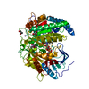
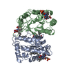
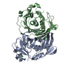
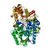
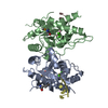
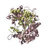
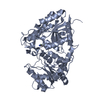
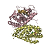
 Rhodospirillum rubrum (bacteria)
 X-RAY DIFFRACTION /
  Authors
 Citation
 Structure visualization
 Downloads & links
 Other downloads

 PDBj
PDBj
 Assembly


 Mass: 55.845 Da / Num. of mol.: 8 / Source method: obtained synthetically / Formula: Fe
 Mass: 54.938 Da / Num. of mol.: 8 / Source method: obtained synthetically / Formula: Mn
 Mass: 35.453 Da / Num. of mol.: 5 / Source method: obtained synthetically / Formula: Cl
 Mass: 62.068 Da / Num. of mol.: 13 / Source method: obtained synthetically / Formula: C2H6O2
 Mass: 59.044 Da / Num. of mol.: 4 / Source method: obtained synthetically / Formula: C2H3O2
 Sample preparation
 / Beamline: BL9-2 / Wavelength: 0.97911
  Processing