 Movie
Movie Controller

[English] 日本語
 Yorodumi
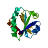
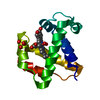
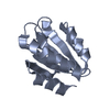
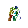
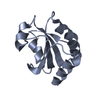
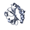
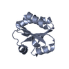
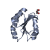
- PDB-2xbi: Crystal structure of Schistosoma mansoni Thioredoxin at 1.6 Angstrom -
+ Open data

- Basic information
| Entry | Database: PDB / ID: 2xbi | ||||||
|---|---|---|---|---|---|---|---|
| Title | Crystal structure of Schistosoma mansoni Thioredoxin at 1.6 Angstrom | ||||||
 Components Components | THIOREDOXIN | ||||||
 Keywords Keywords | OXIDOREDUCTASE / PROTEIN DISULFIDE REDUCTASE | ||||||
| Function / homology |  Function and homology information Function and homology information | ||||||
| Biological species |  | ||||||
| Method |  X-RAY DIFFRACTION / SYNCHROTRON / MOLECULAR REPLACEMENT / Resolution: 1.6 Å X-RAY DIFFRACTION / SYNCHROTRON / MOLECULAR REPLACEMENT / Resolution: 1.6 Å | ||||||
 Authors Authors | Boumis, G. / Miele, A.E. / Dimastrogiovanni, D. / Angelucci, F. / Bellelli, A. | ||||||
 Citation Citation | Journal: Protein Sci. / Year: 2011 Title: Structural and Functional Characterization of Schistosoma Mansoni Thioredoxin. Authors: Boumis, G. / Angelucci, F. / Bellelli, A. / Brunori, M. / Dimastrogiovanni, D. / Miele, A.E. #1: Journal: J.Biol.Chem. / Year: 2010Title: Mapping the Catalytic Cycle of Schistosoma Mansoni Thioredoxin Glutathione Reductase by X-Ray Crystallography. Authors: Angelucci, F. / Dimastrogiovanni, D. / Boumis, G. / Brunori, M. / Miele, A.E. / Saccoccia, F. / Bellelli, A. | ||||||
| History |
|
- Structure visualization
| Structure viewer | Molecule: MolmilJmol/JSmol |
|---|
- Downloads & links
-Download
| PDBx/mmCIF format | 2xbi.cif.gz | 36 KB | Display | PDBx/mmCIF format |
|---|---|---|---|---|
| PDB format | pdb2xbi.ent.gz | 23.6 KB | Display | PDB format |
| PDBx/mmJSON format | 2xbi.json.gz | Tree view | PDBx/mmJSON format | |
| Others |  Other downloads Other downloads |
-Validation report
| Arichive directory | https://data.pdbj.org/pub/pdb/validation_reports/xb/2xbiftp://data.pdbj.org/pub/pdb/validation_reports/xb/2xbi | HTTPS FTP |
|---|
-Related structure data
| Related structure data |  2xbqC  2xc2C  1xwaS S: Starting model for refinement C: citing same article ( |
|---|---|
| Similar structure data |


-Links
 PDBj
PDBj

- Assembly
| Deposited unit | 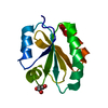
| ||||||||
|---|---|---|---|---|---|---|---|---|---|
| 1 |
| ||||||||
| Unit cell |
|
-Components
| #1: Protein | Mass: 12085.128 Da / Num. of mol.: 1 Source method: isolated from a genetically manipulated source Source: (gene. exp.)  |
|---|---|
| #2: Chemical | ChemComp-GOL /   Mass: 92.094 Da / Num. of mol.: 1 / Source method: obtained synthetically / Formula: C3H8O3 Mass: 92.094 Da / Num. of mol.: 1 / Source method: obtained synthetically / Formula: C3H8O3 |
| #3: Water | ChemComp-HOH /  Mass: 18.015 Da / Num. of mol.: 84 / Source method: isolated from a natural source / Formula: H2O Mass: 18.015 Da / Num. of mol.: 84 / Source method: isolated from a natural source / Formula: H2O |
| Has protein modification | Y |
| Sequence details | GS AT N-TERMINUS COME FROM THROMBIN CLEAVAGE OF THE EXPRESSION |
-Experimental details
-Experiment
| Experiment | Method: X-RAY DIFFRACTION / Number of used crystals: 1 |
|---|
- Sample preparation
| Crystal | Density Matthews: 2.13 Å3/Da / Density % sol: 42.2 % / Description: NONE |
|---|---|
| Crystal grow | pH: 6.5 Details: 0.1M BISTRIS, PH 6.5, 45% POLYPROPYLENE GLYCOL P400 |
-Data collection
| Diffraction | Mean temperature: 100 K |
|---|---|
| Diffraction source | Source: SYNCHROTRON / Site: ESRF  / Beamline: ID14-1 / Wavelength: 0.934 / Beamline: ID14-1 / Wavelength: 0.934 |
| Detector | Type: ADSC CCD / Detector: CCD / Date: Feb 27, 2008 |
| Radiation | Protocol: SINGLE WAVELENGTH / Monochromatic (M) / Laue (L): M / Scattering type: x-ray |
| Radiation wavelength | Wavelength: 0.934 Å / Relative weight: 1 |
| Reflection | Resolution: 1.6→36.01 Å / Num. obs: 14224 / % possible obs: 99.8 % / Observed criterion σ(I): 2 / Redundancy: 6.9 % / Biso Wilson estimate: 9.4 Å2 / Rmerge(I) obs: 0.11 / Net I/σ(I): 14.6 |
| Reflection shell | Resolution: 1.6→1.69 Å / Redundancy: 6.9 % / Rmerge(I) obs: 0.49 / Mean I/σ(I) obs: 4.7 / % possible all: 99.5 |
- Processing
| Software |
| ||||||||||||||||||||||||||||||||||||||||||||||||||||||||||||||||||||||||||||||||||||||||||||||||||||||||||||||||||||||||||||||||||||||||||||||||||||||||||||||||||||||||||||||||||||||
|---|---|---|---|---|---|---|---|---|---|---|---|---|---|---|---|---|---|---|---|---|---|---|---|---|---|---|---|---|---|---|---|---|---|---|---|---|---|---|---|---|---|---|---|---|---|---|---|---|---|---|---|---|---|---|---|---|---|---|---|---|---|---|---|---|---|---|---|---|---|---|---|---|---|---|---|---|---|---|---|---|---|---|---|---|---|---|---|---|---|---|---|---|---|---|---|---|---|---|---|---|---|---|---|---|---|---|---|---|---|---|---|---|---|---|---|---|---|---|---|---|---|---|---|---|---|---|---|---|---|---|---|---|---|---|---|---|---|---|---|---|---|---|---|---|---|---|---|---|---|---|---|---|---|---|---|---|---|---|---|---|---|---|---|---|---|---|---|---|---|---|---|---|---|---|---|---|---|---|---|---|---|---|---|
| Refinement | Method to determine structure: MOLECULAR REPLACEMENT Starting model: PDB ENTRY 1XWA Resolution: 1.6→36.01 Å / Cor.coef. Fo:Fc: 0.939 / Cor.coef. Fo:Fc free: 0.916 / SU B: 1.809 / SU ML: 0.064 / Cross valid method: THROUGHOUT / ESU R: 0.101 / ESU R Free: 0.099 / Stereochemistry target values: MAXIMUM LIKELIHOOD / Details: HYDROGENS HAVE BEEN ADDED IN THE RIDING POSITIONS.
| ||||||||||||||||||||||||||||||||||||||||||||||||||||||||||||||||||||||||||||||||||||||||||||||||||||||||||||||||||||||||||||||||||||||||||||||||||||||||||||||||||||||||||||||||||||||
| Solvent computation | Ion probe radii: 0.8 Å / Shrinkage radii: 0.8 Å / VDW probe radii: 1.2 Å / Solvent model: MASK | ||||||||||||||||||||||||||||||||||||||||||||||||||||||||||||||||||||||||||||||||||||||||||||||||||||||||||||||||||||||||||||||||||||||||||||||||||||||||||||||||||||||||||||||||||||||
| Displacement parameters | Biso mean: 9.734 Å2
| ||||||||||||||||||||||||||||||||||||||||||||||||||||||||||||||||||||||||||||||||||||||||||||||||||||||||||||||||||||||||||||||||||||||||||||||||||||||||||||||||||||||||||||||||||||||
| Refinement step | Cycle: LAST / Resolution: 1.6→36.01 Å
| ||||||||||||||||||||||||||||||||||||||||||||||||||||||||||||||||||||||||||||||||||||||||||||||||||||||||||||||||||||||||||||||||||||||||||||||||||||||||||||||||||||||||||||||||||||||
| Refine LS restraints |
|