 Movie
Movie Controller

[English] 日本語
 Yorodumi
- PDB-1fb6: CRYSTAL STRUCTURE OF THIOREDOXIN M FROM SPINACH CHLOROPLAST (OXID... -
+ Open data

- Basic information
| Entry | Database: PDB / ID: 1fb6 | ||||||
|---|---|---|---|---|---|---|---|
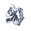
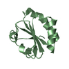
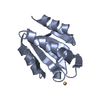
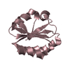
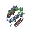
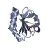
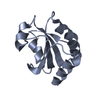
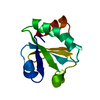
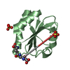
| Title | CRYSTAL STRUCTURE OF THIOREDOXIN M FROM SPINACH CHLOROPLAST (OXIDIZED FORM) | ||||||
 Components Components | THIOREDOXIN M | ||||||
 Keywords Keywords | ELECTRON TRANSPORT | ||||||
| Function / homology |  Function and homology information Function and homology informationprotein-disulfide reductase activity / chloroplast / enzyme activator activity / cytoplasm Similarity search - Function | ||||||
| Biological species |  Spinacia oleracea (spinach) Spinacia oleracea (spinach) | ||||||
| Method |  X-RAY DIFFRACTION / Resolution: 2.1 Å X-RAY DIFFRACTION / Resolution: 2.1 Å | ||||||
 Authors Authors | Capitani, G. / Markovic-Housley, Z. / DelVal, G. / Morris, M. / Jansonius, J.N. / Schurmann, P. | ||||||
 Citation Citation | Journal: J.Mol.Biol. / Year: 2000 Title: Crystal structures of two functionally different thioredoxins in spinach chloroplasts. Authors: Capitani, G. / Markovic-Housley, Z. / DelVal, G. / Morris, M. / Jansonius, J.N. / Schurmann, P. | ||||||
| History |
|
- Structure visualization
| Structure viewer | Molecule: MolmilJmol/JSmol |
|---|
- Downloads & links
-Download
| PDBx/mmCIF format | 1fb6.cif.gz | 53.8 KB | Display | PDBx/mmCIF format |
|---|---|---|---|---|
| PDB format | pdb1fb6.ent.gz | 39.5 KB | Display | PDB format |
| PDBx/mmJSON format | 1fb6.json.gz | Tree view | PDBx/mmJSON format | |
| Others |  Other downloads Other downloads |
-Validation report
| Arichive directory | https://data.pdbj.org/pub/pdb/validation_reports/fb/1fb6ftp://data.pdbj.org/pub/pdb/validation_reports/fb/1fb6 | HTTPS FTP |
|---|
-Related structure data


-Links
 PDBj
PDBj

- Assembly
| Deposited unit | 
| ||||||||
|---|---|---|---|---|---|---|---|---|---|
| 1 | 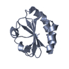
| ||||||||
| 2 | 
| ||||||||
| Unit cell |
| ||||||||
| Details | The biological assembly is a monomer |
-Components
| #1: Protein | Mass: 11793.436 Da / Num. of mol.: 2 / Fragment: OXIDIZED FORM Source method: isolated from a genetically manipulated source Source: (gene. exp.) Spinacia oleracea (spinach) / Cellular location: CHLOROPLAST / Plasmid: PKK233-2 (MODIFIED) / Production host:  #2: Water | ChemComp-HOH / |  Mass: 18.015 Da / Num. of mol.: 121 / Source method: isolated from a natural source / Formula: H2O Mass: 18.015 Da / Num. of mol.: 121 / Source method: isolated from a natural source / Formula: H2OHas protein modification | Y | |
|---|
-Experimental details
-Experiment
| Experiment | Method: X-RAY DIFFRACTION / Number of used crystals: 1 |
|---|
- Sample preparation
| Crystal | Density Matthews: 2.53 Å3/Da / Density % sol: 51.31 % | ||||||||||||||||||||||||||||||||||||||||||
|---|---|---|---|---|---|---|---|---|---|---|---|---|---|---|---|---|---|---|---|---|---|---|---|---|---|---|---|---|---|---|---|---|---|---|---|---|---|---|---|---|---|---|---|
| Crystal grow | Temperature: 293 K / Method: vapor diffusion, hanging drop Details: sodium acetate, PEG monomethylether 2000, ammonium sulphate, VAPOR DIFFUSION, HANGING DROP, temperature 293K | ||||||||||||||||||||||||||||||||||||||||||
| Crystal grow | *PLUS Temperature: 20 ℃ / pH: 7.3 | ||||||||||||||||||||||||||||||||||||||||||
| Components of the solutions | *PLUS
|
-Data collection
| Diffraction | Mean temperature: 277 K |
|---|---|
| Diffraction source | Source: ROTATING ANODE / Type: ELLIOTT GX-20 / Wavelength: 1.5418 |
| Detector | Type: MARRESEARCH / Detector: IMAGE PLATE / Date: Mar 31, 1997 |
| Radiation | Protocol: SINGLE WAVELENGTH / Monochromatic (M) / Laue (L): M / Scattering type: x-ray |
| Radiation wavelength | Wavelength: 1.5418 Å / Relative weight: 1 |
| Reflection | Resolution: 2.1→29.6 Å / Num. obs: 14256 / % possible obs: 99.3 % / Observed criterion σ(I): -3 / Redundancy: 8.5 % / Biso Wilson estimate: 24 Å2 / Rmerge(I) obs: 0.086 / Net I/σ(I): 10.2 |
| Reflection shell | Resolution: 2.1→2.15 Å / Redundancy: 7.4 % / Rmerge(I) obs: 0.396 / % possible all: 92 |
| Reflection shell | *PLUS % possible obs: 92 % |
- Processing
| Software |
| ||||||||||||||||||||
|---|---|---|---|---|---|---|---|---|---|---|---|---|---|---|---|---|---|---|---|---|---|
| Refinement | Resolution: 2.1→29.6 Å / σ(F): 0 / Stereochemistry target values: Engh & Huber Details: Used overall anisotropic B-factor refinement and bulk solvent correction (X-PLOR 3.851 and CNS)
| ||||||||||||||||||||
| Refinement step | Cycle: LAST / Resolution: 2.1→29.6 Å
| ||||||||||||||||||||
| Refine LS restraints |
| ||||||||||||||||||||
| Software | *PLUS Name: X-PLOR / Version: 3.851 / Classification: refinement | ||||||||||||||||||||
| Refinement | *PLUS Highest resolution: 2.1 Å / Lowest resolution: 29.6 Å / σ(F): 0 / % reflection Rfree: 3.8 % / Rfactor obs: 0.204 / Rfactor Rfree: 0.23 | ||||||||||||||||||||
| Solvent computation | *PLUS | ||||||||||||||||||||
| Displacement parameters | *PLUS Biso mean: 21.3 Å2 | ||||||||||||||||||||
| Refine LS restraints | *PLUS
|
