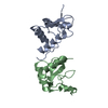
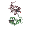
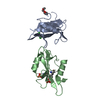
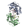
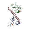
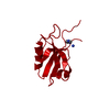
[English] 日本語
Yorodumi
- PDB-2x7z: Crystal Structure of the SAP97 PDZ2 I342W C378A mutant protein domain -

+
Open data


ID or keywords:

Loading...

-
Basic information

Entry
Database: PDB / ID: 2x7z
TitleCrystal Structure of the SAP97 PDZ2 I342W C378A mutant protein domain
ComponentsDISKS LARGE HOMOLOG 1
KeywordsSTRUCTURAL PROTEIN / SH3 DOMAIN / PHOSPHOPROTEIN / SYNAPTIC PROTEIN / HOST-VIRUS INTERACTION
Function / homology
Function and homology information


regulation of voltage-gated potassium channel activity involved in ventricular cardiac muscle cell action potential repolarization / regulation of protein localization to synapse / regulation of potassium ion import / L27 domain binding / regulation of potassium ion export across plasma membrane / MPP7-DLG1-LIN7 complex / membrane raft organization / hard palate development / establishment of centrosome localization / cortical microtubule organization ...regulation of voltage-gated potassium channel activity involved in ventricular cardiac muscle cell action potential repolarization / regulation of protein localization to synapse / regulation of potassium ion import / L27 domain binding / regulation of potassium ion export across plasma membrane / MPP7-DLG1-LIN7 complex / membrane raft organization / hard palate development / establishment of centrosome localization / cortical microtubule organization / negative regulation of p38MAPK cascade / guanylate kinase activity / NrCAM interactions / regulation of sodium ion transmembrane transport / embryonic skeletal system morphogenesis / astral microtubule organization / structural constituent of postsynaptic density / lateral loop / reproductive structure development / myelin sheath abaxonal region / immunological synapse formation / peristalsis / Synaptic adhesion-like molecules / cell projection membrane / smooth muscle tissue development / bicellular tight junction assembly / positive regulation of potassium ion transport / regulation of ventricular cardiac muscle cell action potential / amyloid precursor protein metabolic process / node of Ranvier / Trafficking of AMPA receptors / establishment or maintenance of epithelial cell apical/basal polarity / protein-containing complex localization / Assembly and cell surface presentation of NMDA receptors / endothelial cell proliferation / neurotransmitter receptor localization to postsynaptic specialization membrane / lens development in camera-type eye / regulation of myelination / Activation of Ca-permeable Kainate Receptor / cortical actin cytoskeleton organization / establishment or maintenance of cell polarity / branching involved in ureteric bud morphogenesis / negative regulation of G1/S transition of mitotic cell cycle / positive regulation of actin filament polymerization / receptor clustering / Negative regulation of NMDA receptor-mediated neuronal transmission / Unblocking of NMDA receptors, glutamate binding and activation / phosphoprotein phosphatase activity / Long-term potentiation / immunological synapse / basement membrane / negative regulation of phosphatidylinositol 3-kinase/protein kinase B signal transduction / bicellular tight junction / intercalated disc / potassium channel regulator activity / lateral plasma membrane / phosphatase binding / T cell proliferation / negative regulation of T cell proliferation / actin filament polymerization / Ras activation upon Ca2+ influx through NMDA receptor / regulation of membrane potential / cytoskeletal protein binding / phosphatidylinositol 3-kinase/protein kinase B signal transduction / synaptic membrane / actin filament organization / protein localization to plasma membrane / postsynaptic density membrane / positive regulation of protein localization to plasma membrane / adherens junction / sarcolemma / neuromuscular junction / cytoplasmic side of plasma membrane / negative regulation of ERK1 and ERK2 cascade / cell-cell adhesion / kinase binding / negative regulation of epithelial cell proliferation / cell-cell junction / cell junction / regulation of cell shape / chemical synaptic transmission / RAF/MAP kinase cascade / basolateral plasma membrane / microtubule / transmembrane transporter binding / molecular adaptor activity / neuron projection / cadherin binding / membrane raft / apical plasma membrane / glutamatergic synapse / positive regulation of cell population proliferation / endoplasmic reticulum membrane / perinuclear region of cytoplasm / Golgi apparatus / negative regulation of transcription by RNA polymerase II / endoplasmic reticulum / extracellular exosome / nucleus / plasma membrane
Similarity search - Function
L27-1 / L27_1 / domain in receptor targeting proteins Lin-2 and Lin-7 / L27 domain / L27 domain profile. / Polyubiquitination (PEST) N-terminal domain of MAGUK / L27 domain superfamily / Disks large homologue 1, N-terminal PEST domain / Polyubiquitination (PEST) N-terminal domain of MAGUK / PDZ-associated domain of NMDA receptors ...L27-1 / L27_1 / domain in receptor targeting proteins Lin-2 and Lin-7 / L27 domain / L27 domain profile. / Polyubiquitination (PEST) N-terminal domain of MAGUK / L27 domain superfamily / Disks large homologue 1, N-terminal PEST domain / Polyubiquitination (PEST) N-terminal domain of MAGUK / PDZ-associated domain of NMDA receptors / PDZ-associated domain of NMDA receptors / Disks large 1-like / Guanylate kinase, conserved site / Guanylate kinase-like signature. / Guanylate kinase-like domain profile. / Guanylate kinase-like domain / Guanylate kinase/L-type calcium channel beta subunit / Guanylate kinase / Guanylate kinase homologues. / PDZ domain / Pdz3 Domain / PDZ domain / SH3 domain / PDZ domain profile. / Domain present in PSD-95, Dlg, and ZO-1/2. / PDZ domain / PDZ superfamily / Src homology 3 domains / SH3-like domain superfamily / Src homology 3 (SH3) domain profile. / SH3 domain / Roll / P-loop containing nucleoside triphosphate hydrolase / Mainly Beta
Similarity search - Domain/homology
IMIDAZOLE / AMMONIUM ION / Disks large homolog 1
Similarity search - Component
Biological speciesHOMO SAPIENS (human)
MethodX-RAY DIFFRACTION / SYNCHROTRON / MOLECULAR REPLACEMENT / Resolution: 2 Å
AuthorsHaq, S.R. / Jurgens, M.C. / Chi, C.N. / Elfstrom, L. / Koh, C.S. / Selmer, M. / Gianni, S. / Jemth, P.
CitationJournal: J.Biol.Chem. / Year: 2010
Title: The Plastic Energy Landscape of Protein Folding: A Triangular Folding Mechanism with an Equilibrium Intermediate for a Small Protein Domain.
Authors: Haq, S.R. / Jurgens, M.C. / Chi, C.N. / Elfstrom, L. / Koh, C.S. / Selmer, M. / Gianni, S. / Jemth, P.
History
DepositionMar 4, 2010Deposition site: PDBE / Processing site: PDBE
Revision 1.0Mar 31, 2010Provider: repository / Type: Initial release
Revision 1.1Jul 13, 2011Group: Advisory / Version format compliance
Revision 1.2Jan 17, 2018Group: Data collection / Category: diffrn_source / Item: _diffrn_source.pdbx_synchrotron_site
Revision 1.3Mar 6, 2019Group: Data collection / Experimental preparation / Category: exptl_crystal_grow / Item: _exptl_crystal_grow.method
Revision 1.4Dec 20, 2023Group: Data collection / Database references ...Data collection / Database references / Derived calculations / Other / Refinement description
Category: chem_comp_atom / chem_comp_bond ...chem_comp_atom / chem_comp_bond / database_2 / pdbx_database_status / pdbx_initial_refinement_model / struct_site
Item: _database_2.pdbx_DOI / _database_2.pdbx_database_accession ..._database_2.pdbx_DOI / _database_2.pdbx_database_accession / _pdbx_database_status.status_code_sf / _struct_site.pdbx_auth_asym_id / _struct_site.pdbx_auth_comp_id / _struct_site.pdbx_auth_seq_id

-
Structure visualization

Structure viewerMolecule:
MolmilJmol/JSmol

Downloads & links

-
Assembly

Deposited unit
A: DISKS LARGE HOMOLOG 1
hetero molecules


Theoretical massNumber of molelcules
Total (without water)10,5713
Polymers10,4841
Non-polymers872
Water1,47782
1
A: DISKS LARGE HOMOLOG 1
hetero molecules

A: DISKS LARGE HOMOLOG 1
hetero molecules


Theoretical massNumber of molelcules
Total (without water)21,1426
Polymers20,9682
Non-polymers1744
Water362
TypeNameSymmetry operationNumber
identity operation1_555x,y,z1
crystal symmetry operation5_555-x,y,-z1
Buried area2180 Å2
ΔGint2 kcal/mol
Surface area11230 Å2
MethodPISA
Unit cell
Length a, b, c (Å)47.905, 47.905, 123.287
Angle α, β, γ (deg.)90.00, 90.00, 90.00
Int Tables number95
Space group name H-MP4322

-
Components

#1: Protein DISKS LARGE HOMOLOG 1 / SYNAPSE-ASSOCIATED PROTEIN 97 / SAP97 / SAP-97 / HDLG


Mass: 10484.023 Da / Num. of mol.: 1 / Fragment: PDZ2 DOMAIN, RESIDUES 260-356 / Mutation: YES
Source method: isolated from a genetically manipulated source
Source: (gene. exp.) HOMO SAPIENS (human) / Plasmid: PRSET-SAP97-PDZ2-I342W-C378A / Production host: ESCHERICHIA COLI (E. coli) / Strain (production host): BL21(DE3)PLYSS / References: UniProt: Q12959
#2: Chemical ChemComp-NH4 / AMMONIUM ION / Ammonium


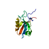
Mass: 18.038 Da / Num. of mol.: 1 / Source method: obtained synthetically / Formula: H4N
#3: Chemical ChemComp-IMD / IMIDAZOLE / Imidazole


Mass: 69.085 Da / Num. of mol.: 1 / Source method: obtained synthetically / Formula: C3H5N2
#4: Water ChemComp-HOH / water / Water


Mass: 18.015 Da / Num. of mol.: 82 / Source method: isolated from a natural source / Formula: H2O

-
Experimental details

-
Experiment

ExperimentMethod: X-RAY DIFFRACTION / Number of used crystals: 1

-
Sample preparation

CrystalDensity Matthews: 3.37 Å3/Da / Density % sol: 63.5 % / Description: NONE
Crystal growMethod: vapor diffusion / pH: 8.4
Details: VAPOR DIFFUSION, 100MM TRIS-HCL PH 8.4, 2.4M AMMONIUM SULPHATE

-
Data collection

DiffractionMean temperature: 100 K
Diffraction sourceSource: SYNCHROTRON / Site: MAX II / Beamline: I911-2 / Wavelength: 1.04
DetectorType: MARRESEARCH SX-165 / Detector: CCD / Date: Jan 21, 2009 / Details: MIRRORS
RadiationMonochromator: SILICON CRYSTAL / Protocol: SINGLE WAVELENGTH / Monochromatic (M) / Laue (L): M / Scattering type: x-ray
Radiation wavelengthWavelength: 1.04 Å / Relative weight: 1
ReflectionResolution: 2→29.9 Å / Num. obs: 10153 / % possible obs: 96.6 % / Observed criterion σ(I): 0 / Redundancy: 20.4 % / Rmerge(I) obs: 0.08 / Net I/σ(I): 31.5
Reflection shellResolution: 2→2.05 Å / Redundancy: 20.8 % / Rmerge(I) obs: 0.22 / Mean I/σ(I) obs: 15.4 / % possible all: 96.8

-
Processing

Software
NameVersionClassification
REFMAC5.5.0102refinement
XDSdata reduction
XSCALEdata scaling
PHASERphasing
RefinementMethod to determine structure: MOLECULAR REPLACEMENT
Starting model: PDB ENTRY 2AWU
Resolution: 2→29.69 Å / Cor.coef. Fo:Fc: 0.947 / Cor.coef. Fo:Fc free: 0.921 / SU B: 7.174 / SU ML: 0.092 / TLS residual ADP flag: LIKELY RESIDUAL / Cross valid method: THROUGHOUT / ESU R: 0.141 / ESU R Free: 0.137 / Stereochemistry target values: MAXIMUM LIKELIHOOD
Details: HYDROGENS HAVE BEEN ADDED IN THE RIDING POSITIONS ATOM RECORD CONTAINS RESIDUAL B FACTORS ONLY
RfactorNum. reflection% reflectionSelection details
Rfree0.23236 516 5 %RANDOM
Rwork0.19381 ---
obs0.19576 9801 100 %-
Solvent computationIon probe radii: 0.8 Å / Shrinkage radii: 0.8 Å / VDW probe radii: 1.4 Å / Solvent model: MASK
Displacement parametersBiso mean: 16.951 Å2
Baniso -1Baniso -2Baniso -3
1-0.1 Å20 Å20 Å2
2--0.1 Å20 Å2
3----0.2 Å2
Refinement stepCycle: LAST / Resolution: 2→29.69 Å
ProteinNucleic acidLigandSolventTotal
Num. atoms738 0 6 82 826
Refine LS restraints
Refine-IDTypeDev idealDev ideal targetNumber
X-RAY DIFFRACTIONr_bond_refined_d0.0220.022755
X-RAY DIFFRACTIONr_bond_other_d
X-RAY DIFFRACTIONr_angle_refined_deg1.6911.9821017
X-RAY DIFFRACTIONr_angle_other_deg
X-RAY DIFFRACTIONr_dihedral_angle_1_deg6.556598
X-RAY DIFFRACTIONr_dihedral_angle_2_deg38.52826.92326
X-RAY DIFFRACTIONr_dihedral_angle_3_deg15.15415138
X-RAY DIFFRACTIONr_dihedral_angle_4_deg
X-RAY DIFFRACTIONr_chiral_restr0.1320.2117
X-RAY DIFFRACTIONr_gen_planes_refined0.0090.021547
X-RAY DIFFRACTIONr_gen_planes_other
X-RAY DIFFRACTIONr_nbd_refined
X-RAY DIFFRACTIONr_nbd_other
X-RAY DIFFRACTIONr_nbtor_refined
X-RAY DIFFRACTIONr_nbtor_other
X-RAY DIFFRACTIONr_xyhbond_nbd_refined
X-RAY DIFFRACTIONr_xyhbond_nbd_other
X-RAY DIFFRACTIONr_metal_ion_refined
X-RAY DIFFRACTIONr_metal_ion_other
X-RAY DIFFRACTIONr_symmetry_vdw_refined
X-RAY DIFFRACTIONr_symmetry_vdw_other
X-RAY DIFFRACTIONr_symmetry_hbond_refined
X-RAY DIFFRACTIONr_symmetry_hbond_other
X-RAY DIFFRACTIONr_symmetry_metal_ion_refined
X-RAY DIFFRACTIONr_symmetry_metal_ion_other
X-RAY DIFFRACTIONr_mcbond_it1.0441.5487
X-RAY DIFFRACTIONr_mcbond_other
X-RAY DIFFRACTIONr_mcangle_it1.7642781
X-RAY DIFFRACTIONr_mcangle_other
X-RAY DIFFRACTIONr_scbond_it2.9993268
X-RAY DIFFRACTIONr_scbond_other
X-RAY DIFFRACTIONr_scangle_it5.0564.5236
X-RAY DIFFRACTIONr_scangle_other
X-RAY DIFFRACTIONr_long_range_B_refined
X-RAY DIFFRACTIONr_long_range_B_other
X-RAY DIFFRACTIONr_rigid_bond_restr
X-RAY DIFFRACTIONr_sphericity_free
X-RAY DIFFRACTIONr_sphericity_bonded
LS refinement shellResolution: 2→2.052 Å / Total num. of bins used: 20
RfactorNum. reflection% reflection
Rfree0.287 36 -
Rwork0.212 686 -
obs--100 %
Refinement TLS params.Method: refined / Origin x: 10.8916 Å / Origin y: -4.2617 Å / Origin z: 10.8723 Å
111213212223313233
T0.0267 Å20.0149 Å2-0.003 Å2-0.0782 Å20.0209 Å2--0.0717 Å2
L4.3035 °2-0.5061 °2-0.8472 °2-1.3123 °20.6062 °2--4.6422 °2
S-0.0756 Å °-0.0212 Å °-0.0649 Å °0.0685 Å °0.0399 Å °-0.2384 Å °0.2071 Å °0.534 Å °0.0357 Å °

+
About Yorodumi

-
News

-
Feb 9, 2022. New format data for meta-information of EMDB entries

New format data for meta-information of EMDB entries

  • Version 3 of the EMDB header file is now the official format.
  • The previous official version 1.9 will be removed from the archive.

Related info.:EMDB header

External links:wwPDB to switch to version 3 of the EMDB data model

-
Aug 12, 2020. Covid-19 info

Covid-19 info

URL: https://pdbj.org/emnavi/covid19.php

New page: Covid-19 featured information page in EM Navigator.

Related info.:Covid-19 info / Mar 5, 2020. Novel coronavirus structure data

+
Mar 5, 2020. Novel coronavirus structure data

Novel coronavirus structure data

Related info.:Yorodumi Speices / Aug 12, 2020. Covid-19 info

External links:COVID-19 featured content - PDBj / Molecule of the Month (242):Coronavirus Proteases

+
Jan 31, 2019. EMDB accession codes are about to change! (news from PDBe EMDB page)

EMDB accession codes are about to change! (news from PDBe EMDB page)

  • The allocation of 4 digits for EMDB accession codes will soon come to an end. Whilst these codes will remain in use, new EMDB accession codes will include an additional digit and will expand incrementally as the available range of codes is exhausted. The current 4-digit format prefixed with “EMD-” (i.e. EMD-XXXX) will advance to a 5-digit format (i.e. EMD-XXXXX), and so on. It is currently estimated that the 4-digit codes will be depleted around Spring 2019, at which point the 5-digit format will come into force.
  • The EM Navigator/Yorodumi systems omit the EMD- prefix.

Related info.:Q: What is EMD? / ID/Accession-code notation in Yorodumi/EM Navigator

External links:EMDB Accession Codes are Changing Soon! / Contact to PDBj

+
Jul 12, 2017. Major update of PDB

Major update of PDB

  • wwPDB released updated PDB data conforming to the new PDBx/mmCIF dictionary.
  • This is a major update changing the version number from 4 to 5, and with Remediation, in which all the entries are updated.
  • In this update, many items about electron microscopy experimental information are reorganized (e.g. em_software).
  • Now, EM Navigator and Yorodumi are based on the updated data.

External links:wwPDB Remediation / Enriched Model Files Conforming to OneDep Data Standards Now Available in the PDB FTP Archive

-
Yorodumi

Thousand views of thousand structures

  • Yorodumi is a browser for structure data from EMDB, PDB, SASBDB, etc.
  • This page is also the successor to EM Navigator detail page, and also detail information page/front-end page for Omokage search.
  • The word "yorodu" (or yorozu) is an old Japanese word meaning "ten thousand". "mi" (miru) is to see.

Related info.:EMDB / PDB / SASBDB / Comparison of 3 databanks / Yorodumi Search / Aug 31, 2016. New EM Navigator & Yorodumi / Yorodumi Papers / Jmol/JSmol / Function and homology information / Changes in new EM Navigator and Yorodumi

Read more