 Movie
Movie Controller

[English] 日本語
 Yorodumi
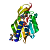
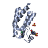
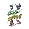
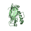
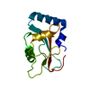
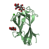
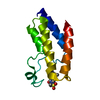
- PDB-1m07: RESIDUES INVOLVED IN THE CATALYSIS AND BASE SPECIFICITY OF CYTOTO... -
+ Open data

- Basic information
| Entry | Database: PDB / ID: 1m07 | |||||||||
|---|---|---|---|---|---|---|---|---|---|---|
| Title | RESIDUES INVOLVED IN THE CATALYSIS AND BASE SPECIFICITY OF CYTOTOXIC RIBONUCLEASE FROM BULLFROG (RANA CATESBEIANA) | |||||||||
 Components Components |
| |||||||||
 Keywords Keywords | HYDROLASE/DNA / RC-RNase-d(ACGA) / ribonuclease / bullfrog / cytotoxicity / HYDROLASE-DNA COMPLEX | |||||||||
| Function / homology |  Function and homology information Function and homology informationHydrolases; Acting on ester bonds; Endoribonucleases producing 3'-phosphomonoesters / RNA nuclease activity / carbohydrate binding / endonuclease activity / nucleic acid binding / defense response to Gram-positive bacterium / hydrolase activity / extracellular region Similarity search - Function | |||||||||
| Biological species | Rana catesbeiana (American bullfrog) | |||||||||
| Method |  X-RAY DIFFRACTION / SYNCHROTRON / MOLECULAR REPLACEMENT / Resolution: 1.8 Å X-RAY DIFFRACTION / SYNCHROTRON / MOLECULAR REPLACEMENT / Resolution: 1.8 Å | |||||||||
 Authors Authors | Leu, Y.-J. / Chern, S.-S. / Wang, S.-C. / Hsiao, Y.-Y. / Amiraslanov, I. / Liaw, Y.-C. / Liao, Y.-D. | |||||||||
 Citation Citation | Journal: J.Biol.Chem. / Year: 2003 Title: Residues involved in the catalysis, base specificity, and cytotoxicity of ribonuclease from Rana catesbeiana based upon mutagenesis and X-ray crystallography Authors: Leu, Y.-J. / Chern, S.-S. / Wang, S.-C. / Hsiao, Y.-Y. / Amiraslanov, I. / Liaw, Y.-C. / Liao, Y.-D. #1: Journal: To be PublishedTitle: THE CRYSTAL STRUCTURE OF A CYTOTOXIC RIBONUCLEASE FROM THE OOCYTE OF RANA CATESBEIANA Authors: Leu, Y.-J. / Chern, S.-S. / Wang, S.-C. / Hsiao, Y.-Y. / Amiraslanov, I.R. / Liaw, Y.-C. / Liao, Y.-D. #2: Journal: J.BIOMOL.NMR / Year: 1996Title: THE SECONDARY STRUCTURE OF A PYRIMIDINE-GUANINE SEQUENCE-SPECIFIC RIBONUCLEASE POSSESSING CYTOTOXIC ACTIVITY FROM THE OOCYTE OF RANA CATESBEIANA Authors: Chen, C. / Hom, K. / Huang, R.F. / Chou, P.J. / Liao, Y.D. / Huang, T. #3: Journal: J.Mol.Biol. / Year: 1998Title: THE SOLUTION STRUCTURE OF A CYTOTOXIC RIBONUCLEASE FROM THE OOCYTE OF RANA CATESBEIANA (BULLFROG) Authors: Chang, C.F. / Chen, C. / Chen, Y.C. / Hom, K. / Huang, R.F. / Huang, T.H. #4: Journal: J.Biol.Chem. / Year: 1998Title: THE RANA CATESBEIANA RCR GENE ENCODING A CYTOTOXIC RIBONUCLEASE : TISSUE DISTRIBUTION, CLONING, PURIFICATION, CYTOTOXICITY, AND ACTIVE RESIDUES FOR RNASE ACTIVITY Authors: Huang, H.C. / Wang, S.C. / Leu, Y.J. / Lu, S.C. / Liao, Y.D. #5: Journal: Nucleic Acids Res. / Year: 2000Title: PURIFICATION AND CLONING OF CYTOTOXIC RIBONUCLEASE FROM RANA CATESBEIANA (BULLFROG) Authors: Liao, Y.D. / Huang, H.C. / Leu, Y.J. / Wei, C.W. / Tang, P.C. / Wang, S.C. | |||||||||
| History |
| |||||||||
| Remark 400 | COMPOUND THE ATOMS (C6, N9, C4, C4', C5', C2', N7, C8, N1) OF RESIDUE 4 CHAIN C,D WAS FIXED IN THE REFINEMENT. |
- Structure visualization
| Structure viewer | Molecule: MolmilJmol/JSmol |
|---|
- Downloads & links
-Download
| PDBx/mmCIF format | 1m07.cif.gz | 72.6 KB | Display | PDBx/mmCIF format |
|---|---|---|---|---|
| PDB format | pdb1m07.ent.gz | 51.7 KB | Display | PDB format |
| PDBx/mmJSON format | 1m07.json.gz | Tree view | PDBx/mmJSON format | |
| Others |  Other downloads Other downloads |
-Validation report
| Arichive directory | https://data.pdbj.org/pub/pdb/validation_reports/m0/1m07ftp://data.pdbj.org/pub/pdb/validation_reports/m0/1m07 | HTTPS FTP |
|---|
-Related structure data
| Related structure data |  1km8S S: Starting model for refinement |
|---|---|
| Similar structure data |


-Links
 PDBj
PDBj

- Assembly
| Deposited unit | 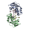
| ||||||||
|---|---|---|---|---|---|---|---|---|---|
| 1 | 
| ||||||||
| 2 | 
| ||||||||
| Unit cell |
|
-Components
| #1: DNA chain | Mass: 1199.844 Da / Num. of mol.: 2 / Source method: obtained synthetically #2: Protein | Mass: 12459.343 Da / Num. of mol.: 2 / Source method: isolated from a natural source / Source: (natural) Rana catesbeiana (American bullfrog) / Organ: oocytes / References: UniProt: P11916, EC: 3.1.27.5 #3: Water | ChemComp-HOH / |  Mass: 18.015 Da / Num. of mol.: 323 / Source method: isolated from a natural source / Formula: H2O Mass: 18.015 Da / Num. of mol.: 323 / Source method: isolated from a natural source / Formula: H2OHas protein modification | Y | |
|---|
-Experimental details
-Experiment
| Experiment | Method: X-RAY DIFFRACTION / Number of used crystals: 1 |
|---|
- Sample preparation
| Crystal | Density Matthews: 2.02 Å3/Da / Density % sol: 38.66 % | |||||||||||||||||||||||||||||||||||||||||||||||||
|---|---|---|---|---|---|---|---|---|---|---|---|---|---|---|---|---|---|---|---|---|---|---|---|---|---|---|---|---|---|---|---|---|---|---|---|---|---|---|---|---|---|---|---|---|---|---|---|---|---|---|
| Crystal grow | Temperature: 298 K / Method: vapor diffusion, hanging drop / pH: 5.6 Details: 0.1 M potassium sodium tartrate tetrahydrate, 24 % PEG 8000, 0.05 M sodium citrate, pH 5.6, VAPOR DIFFUSION, HANGING DROP, temperature 298K | |||||||||||||||||||||||||||||||||||||||||||||||||
| Crystal grow | *PLUS pH: 8 | |||||||||||||||||||||||||||||||||||||||||||||||||
| Components of the solutions | *PLUS
|
-Data collection
| Diffraction | Mean temperature: 298 K |
|---|---|
| Diffraction source | Source: SYNCHROTRON / Site: NSLS  / Beamline: X4A / Wavelength: 1.05 Å / Beamline: X4A / Wavelength: 1.05 Å |
| Detector | Type: ADSC QUANTUM 4 / Detector: CCD / Date: Aug 6, 2000 / Details: KOHZU double crystal monochromator |
| Radiation | Monochromator: SAGITALLY FOCUSED Si(111) / Protocol: SINGLE WAVELENGTH / Monochromatic (M) / Laue (L): M / Scattering type: x-ray |
| Radiation wavelength | Wavelength: 1.05 Å / Relative weight: 1 |
| Reflection | Resolution: 1.79→19.68 Å / Num. all: 20226 / Num. obs: 18075 / % possible obs: 90.1 % / Observed criterion σ(F): 0 / Observed criterion σ(I): 0 / Redundancy: 6.81 % / Biso Wilson estimate: 14.8 Å2 / Rmerge(I) obs: 0.073 / Rsym value: 0.073 / Net I/σ(I): 11.1 |
| Reflection shell | Resolution: 1.8→1.83 Å / Redundancy: 2.1 % / Rmerge(I) obs: 0.18 / Mean I/σ(I) obs: 6.2 / Num. unique all: 894 / Rsym value: 0.18 / % possible all: 90.5 |
| Reflection | *PLUS Highest resolution: 1.8 Å / Num. obs: 20226 / Num. measured all: 137742 |
| Reflection shell | *PLUS % possible obs: 90.5 % / Rmerge(I) obs: 0.18 |
- Processing
| Software |
| ||||||||||||||||||||||||||||||||||||||||||||||||||||||||||||||||||||||||
|---|---|---|---|---|---|---|---|---|---|---|---|---|---|---|---|---|---|---|---|---|---|---|---|---|---|---|---|---|---|---|---|---|---|---|---|---|---|---|---|---|---|---|---|---|---|---|---|---|---|---|---|---|---|---|---|---|---|---|---|---|---|---|---|---|---|---|---|---|---|---|---|---|---|
| Refinement | Method to determine structure: MOLECULAR REPLACEMENT Starting model: PDB ENTRY 1KM8 Resolution: 1.8→19.68 Å / Rfactor Rfree error: 0.006 / Occupancy max: 1 / Occupancy min: 0.5 / Isotropic thermal model: Overall / Cross valid method: THROUGHOUT / σ(F): 0 / σ(I): 0 / Stereochemistry target values: Engh & Huber
| ||||||||||||||||||||||||||||||||||||||||||||||||||||||||||||||||||||||||
| Solvent computation | Solvent model: CNS bulk solvent model used / Bsol: 68.8524 Å2 / ksol: 0.437359 e/Å3 | ||||||||||||||||||||||||||||||||||||||||||||||||||||||||||||||||||||||||
| Displacement parameters | Biso mean: 25.29 Å2
| ||||||||||||||||||||||||||||||||||||||||||||||||||||||||||||||||||||||||
| Refine analyze |
| ||||||||||||||||||||||||||||||||||||||||||||||||||||||||||||||||||||||||
| Refinement step | Cycle: LAST / Resolution: 1.8→19.68 Å
| ||||||||||||||||||||||||||||||||||||||||||||||||||||||||||||||||||||||||
| Refine LS restraints |
| ||||||||||||||||||||||||||||||||||||||||||||||||||||||||||||||||||||||||
| LS refinement shell | Refine-ID: X-RAY DIFFRACTION / Total num. of bins used: 8
| ||||||||||||||||||||||||||||||||||||||||||||||||||||||||||||||||||||||||
| Xplor file |
| ||||||||||||||||||||||||||||||||||||||||||||||||||||||||||||||||||||||||
| Software | *PLUS Name: CNS / Classification: refinement | ||||||||||||||||||||||||||||||||||||||||||||||||||||||||||||||||||||||||
| Refinement | *PLUS Rfactor Rfree: 0.229 | ||||||||||||||||||||||||||||||||||||||||||||||||||||||||||||||||||||||||
| Solvent computation | *PLUS | ||||||||||||||||||||||||||||||||||||||||||||||||||||||||||||||||||||||||
| Displacement parameters | *PLUS | ||||||||||||||||||||||||||||||||||||||||||||||||||||||||||||||||||||||||
| Refine LS restraints | *PLUS
|
