 Movie
Movie Controller

[English] 日本語
 Yorodumi
- PDB-2x2k: Crystal Structure of phosphorylated RET tyrosine kinase domain wi... -
+ Open data

- Basic information
| Entry | Database: PDB / ID: 2x2k | ||||||
|---|---|---|---|---|---|---|---|
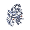
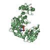
| Title | Crystal Structure of phosphorylated RET tyrosine kinase domain with inhibitor | ||||||
 Components Components | PROTO-ONCOGENE TYROSINE-PROTEIN KINASE RECEPTOR RET | ||||||
 Keywords Keywords | TRANSFERASE / TYROSINE KINASE / HIRSCHSPRUNG DISEASE / TYROSINE-PROTEIN KINASE / PROTO-ONCOGENE / PHOSPHOPROTEIN / PHOSPHOTRANSFERASE | ||||||
| Function / homology |  Function and homology information Function and homology informationPeyer's patch morphogenesis / GDF15-GFRAL signaling pathway / positive regulation of metanephric glomerulus development / ureter maturation / embryonic epithelial tube formation / glial cell-derived neurotrophic factor receptor signaling pathway / lymphocyte migration into lymphoid organs / posterior midgut development / Formation of the ureteric bud / membrane protein proteolysis ...Peyer's patch morphogenesis / GDF15-GFRAL signaling pathway / positive regulation of metanephric glomerulus development / ureter maturation / embryonic epithelial tube formation / glial cell-derived neurotrophic factor receptor signaling pathway / lymphocyte migration into lymphoid organs / posterior midgut development / Formation of the ureteric bud / membrane protein proteolysis / Formation of the nephric duct / enteric nervous system development / neuron cell-cell adhesion / plasma membrane protein complex / neuron maturation / positive regulation of extrinsic apoptotic signaling pathway in absence of ligand / positive regulation of cell adhesion mediated by integrin / neural crest cell migration / ureteric bud development / regulation of axonogenesis / response to pain / homophilic cell-cell adhesion / RET signaling / positive regulation of cell size / regulation of cell adhesion / cellular response to retinoic acid / NPAS4 regulates expression of target genes / transmembrane receptor protein tyrosine kinase activity / axon guidance / cell surface receptor protein tyrosine kinase signaling pathway / positive regulation of neuron projection development / receptor protein-tyrosine kinase / MAPK cascade / signaling receptor activity / RAF/MAP kinase cascade / protein tyrosine kinase activity / positive regulation of phosphatidylinositol 3-kinase/protein kinase B signal transduction / receptor complex / endosome membrane / positive regulation of MAPK cascade / positive regulation of cell migration / axon / calcium ion binding / positive regulation of gene expression / positive regulation of DNA-templated transcription / signal transduction / ATP binding / plasma membrane Similarity search - Function | ||||||
| Biological species |  HOMO SAPIENS (human) HOMO SAPIENS (human) | ||||||
| Method |  X-RAY DIFFRACTION / MOLECULAR REPLACEMENT / Resolution: 2.6 Å X-RAY DIFFRACTION / MOLECULAR REPLACEMENT / Resolution: 2.6 Å | ||||||
 Authors Authors | Knowles, P.P. / Murray-Rust, J. / Kjaer, S. / McDonald, N.Q. | ||||||
 Citation Citation | Journal: Bioorg. Med. Chem. / Year: 2010 Title: Synthesis, structure-activity relationship and crystallographic studies of 3-substituted indolin-2-one RET inhibitors. Authors: Mologni, L. / Rostagno, R. / Brussolo, S. / Knowles, P.P. / Kjaer, S. / Murray-Rust, J. / Rosso, E. / Zambon, A. / Scapozza, L. / McDonald, N.Q. / Lucchini, V. / Gambacorti-Passerini, C. | ||||||
| History |
|
- Structure visualization
| Structure viewer | Molecule: MolmilJmol/JSmol |
|---|
- Downloads & links
-Download
| PDBx/mmCIF format | 2x2k.cif.gz | 73.2 KB | Display | PDBx/mmCIF format |
|---|---|---|---|---|
| PDB format | pdb2x2k.ent.gz | 52.3 KB | Display | PDB format |
| PDBx/mmJSON format | 2x2k.json.gz | Tree view | PDBx/mmJSON format | |
| Others |  Other downloads Other downloads |
-Validation report
| Summary document | 2x2k_validation.pdf.gz | 698.7 KB | Display | wwPDB validaton report |
|---|---|---|---|---|
| Full document | 2x2k_full_validation.pdf.gz | 701.1 KB | Display | |
| Data in XML | 2x2k_validation.xml.gz | 12.8 KB | Display | |
| Data in CIF | 2x2k_validation.cif.gz | 17 KB | Display | |
| Arichive directory | https://data.pdbj.org/pub/pdb/validation_reports/x2/2x2kftp://data.pdbj.org/pub/pdb/validation_reports/x2/2x2k | HTTPS FTP |
-Related structure data
| Related structure data | 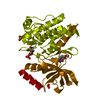 2x2lC  2x2mC 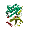 2ivtS S: Starting model for refinement C: citing same article ( |
|---|---|
| Similar structure data |


-Links
 PDBj
PDBj


- Assembly
| Deposited unit | 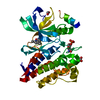
| ||||||||
|---|---|---|---|---|---|---|---|---|---|
| 1 | 
| ||||||||
| Unit cell |
|
-Components
| #1: Protein | Mass: 35788.215 Da / Num. of mol.: 1 / Fragment: TYROSINE KINASE DOMAIN, RESIDUES 705-1013 Source method: isolated from a genetically manipulated source Details: RESIDUES 705-1013,5 N-TERMINAL VECTOR-DERIVED RESIDUES GPLSL Source: (gene. exp.) HOMO SAPIENS (human) / Plasmid: PBACPAK-HIS3 (CLONTECH) MODIFIED / Cell line (production host): SF9 / Production host:   SPODOPTERA FRUGIPERDA (fall armyworm) SPODOPTERA FRUGIPERDA (fall armyworm)References: UniProt: P07949, receptor protein-tyrosine kinase | ||||||||
|---|---|---|---|---|---|---|---|---|---|
| #2: Chemical |   Mass: 46.025 Da / Num. of mol.: 3 / Source method: obtained synthetically / Formula: CH2O2 Mass: 46.025 Da / Num. of mol.: 3 / Source method: obtained synthetically / Formula: CH2O2#3: Chemical | ChemComp-X2K / ( |   Mass: 253.299 Da / Num. of mol.: 1 / Source method: obtained synthetically / Formula: C15H15N3O Mass: 253.299 Da / Num. of mol.: 1 / Source method: obtained synthetically / Formula: C15H15N3O#4: Water | ChemComp-HOH / |  Mass: 18.015 Da / Num. of mol.: 28 / Source method: isolated from a natural source / Formula: H2O Mass: 18.015 Da / Num. of mol.: 28 / Source method: isolated from a natural source / Formula: H2OHas protein modification | Y | Sequence details | 705-1013 CORRESPOND | |
-Experimental details
-Experiment
| Experiment | Method: X-RAY DIFFRACTION / Number of used crystals: 1 |
|---|
- Sample preparation
| Crystal | Density Matthews: 2.9 Å3/Da / Density % sol: 57.7 % / Description: NONE |
|---|---|
| Crystal grow | Temperature: 289 K / Method: vapor diffusion, sitting drop Details: PROTEIN 4.5 MG/ML IN 20 MM TRIS-HCL PH 8, 100MM NACL,1MM DTT, 1MM EDTA RESERVOIR 1.85 M SODIUM FORMATE, 0.1 SODIUM CITRATE PH 5.5, 0.2M LITHIUM CHLORIDE VAPOUR DIFFUSION, SITTING DROP, 289 K |
-Data collection
| Diffraction | Mean temperature: 100 K |
|---|---|
| Diffraction source | Source: ROTATING ANODE / Type: RIGAKU MICROMAX-007 HF / Wavelength: 1.5418 |
| Detector | Type: MARRESEARCH / Detector: IMAGE PLATE / Date: Jul 29, 2007 / Details: MIRRORS |
| Radiation | Protocol: SINGLE WAVELENGTH / Monochromatic (M) / Laue (L): M / Scattering type: x-ray |
| Radiation wavelength | Wavelength: 1.5418 Å / Relative weight: 1 |
| Reflection | Resolution: 2.6→21.64 Å / Num. obs: 11886 / % possible obs: 99.1 % / Observed criterion σ(I): 0 / Redundancy: 3.1 % / Rmerge(I) obs: 0.06 / Net I/σ(I): 17 |
| Reflection shell | Resolution: 2.6→2.74 Å / Redundancy: 3 % / Rmerge(I) obs: 0.23 / Mean I/σ(I) obs: 5.2 / % possible all: 100 |
- Processing
| Software |
| ||||||||||||||||||||||||||||||||||||||||||||||||||||||||||||||||||||||||||||||||||||||||||||||||||||||||||||||||||||||||||||||||||||||||||||||||||||||||||||||||||||||||||||||||||||||
|---|---|---|---|---|---|---|---|---|---|---|---|---|---|---|---|---|---|---|---|---|---|---|---|---|---|---|---|---|---|---|---|---|---|---|---|---|---|---|---|---|---|---|---|---|---|---|---|---|---|---|---|---|---|---|---|---|---|---|---|---|---|---|---|---|---|---|---|---|---|---|---|---|---|---|---|---|---|---|---|---|---|---|---|---|---|---|---|---|---|---|---|---|---|---|---|---|---|---|---|---|---|---|---|---|---|---|---|---|---|---|---|---|---|---|---|---|---|---|---|---|---|---|---|---|---|---|---|---|---|---|---|---|---|---|---|---|---|---|---|---|---|---|---|---|---|---|---|---|---|---|---|---|---|---|---|---|---|---|---|---|---|---|---|---|---|---|---|---|---|---|---|---|---|---|---|---|---|---|---|---|---|---|---|
| Refinement | Method to determine structure: MOLECULAR REPLACEMENT Starting model: PDB ENTRY 2IVT, FLEXIBLE LOOPS REMOVED Resolution: 2.6→30 Å / Cor.coef. Fo:Fc: 0.936 / Cor.coef. Fo:Fc free: 0.884 / SU B: 10.037 / SU ML: 0.216 / Cross valid method: THROUGHOUT / ESU R: 0.524 / ESU R Free: 0.306 / Stereochemistry target values: MAXIMUM LIKELIHOOD / Details: HYDROGENS HAVE BEEN ADDED IN THE RIDING POSITIONS
| ||||||||||||||||||||||||||||||||||||||||||||||||||||||||||||||||||||||||||||||||||||||||||||||||||||||||||||||||||||||||||||||||||||||||||||||||||||||||||||||||||||||||||||||||||||||
| Solvent computation | Ion probe radii: 0.8 Å / Shrinkage radii: 0.8 Å / VDW probe radii: 1.2 Å / Solvent model: BABINET MODEL WITH MASK | ||||||||||||||||||||||||||||||||||||||||||||||||||||||||||||||||||||||||||||||||||||||||||||||||||||||||||||||||||||||||||||||||||||||||||||||||||||||||||||||||||||||||||||||||||||||
| Displacement parameters | Biso mean: 39.5 Å2
| ||||||||||||||||||||||||||||||||||||||||||||||||||||||||||||||||||||||||||||||||||||||||||||||||||||||||||||||||||||||||||||||||||||||||||||||||||||||||||||||||||||||||||||||||||||||
| Refinement step | Cycle: LAST / Resolution: 2.6→30 Å
| ||||||||||||||||||||||||||||||||||||||||||||||||||||||||||||||||||||||||||||||||||||||||||||||||||||||||||||||||||||||||||||||||||||||||||||||||||||||||||||||||||||||||||||||||||||||
| Refine LS restraints |
|