 Movie
Movie Controller

[English] 日本語
 Yorodumi
- PDB-3pmg: STRUCTURE OF RABBIT MUSCLE PHOSPHOGLUCOMUTASE AT 2.4 ANGSTROMS RE... -
+ Open data

- Basic information
| Entry | Database: PDB / ID: 3pmg | |||||||||
|---|---|---|---|---|---|---|---|---|---|---|
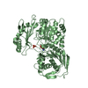
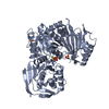
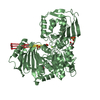
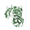
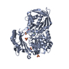
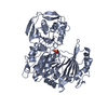
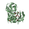
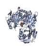
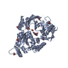
| Title | STRUCTURE OF RABBIT MUSCLE PHOSPHOGLUCOMUTASE AT 2.4 ANGSTROMS RESOLUTION. USE OF FREEZING POINT DEPRESSANT AND REDUCED TEMPERATURE TO ENHANCE DIFFRACTIVITY | |||||||||
 Components Components | Phosphoglucomutase-1 | |||||||||
 Keywords Keywords | ISOMERASE / PHOSPHOTRANSFERASE | |||||||||
| Function / homology |  Function and homology information Function and homology informationphosphoglucomutase (alpha-D-glucose-1,6-bisphosphate-dependent) / phosphoglucomutase activity / sarcoplasmic reticulum / glucose metabolic process / magnesium ion binding / cytosol Similarity search - Function | |||||||||
| Biological species |  | |||||||||
| Method |  X-RAY DIFFRACTION / Resolution: 2.4 Å X-RAY DIFFRACTION / Resolution: 2.4 Å | |||||||||
 Authors Authors | Ray Junior, W.J. / Liu, Y. / Baranidharan, S. | |||||||||
 Citation Citation | Journal: Acta Crystallogr.,Sect.D / Year: 1997 Title: Structure of rabbit muscle phosphoglucomutase refined at 2.4 A resolution. Authors: Liu, Y. / Ray, W.J. / Baranidharan, S. #1: Journal: Biochemistry / Year: 1993Title: Structural Changes at the Metal Ion Binding Site During the Phosphoglucomutase Reaction Authors: Ray Junior, W.J. / Post, C.B. / Liu, Y. / Rhyu, G.I. #2: Journal: J.Biol.Chem. / Year: 1992Title: The Crystal Structure of Phosphoglucomutase Refined at 2.7 Angstroms Resolution Authors: Dai, J.B. / Liu, Y. / Ray, W.J. / Konno, M. #3: Journal: J.Biol.Chem. / Year: 1986Title: The Catalytic Activity of Muscle Phosphoglucomutase in the Crystalline Phase Authors: Ray Junior, W.J. #4: Journal: J.Biol.Chem. / Year: 1986Title: The Structure of Rabbit Muscle Phosphoglucomutase at Intermediate Resolution Authors: Lin, Z.-J. / Konno, M. / Abad-Zapatero, C. / Wierenga, R. / Murthy, M.R.N. / Ray Junior, W.J. / Rossmann, M.G. | |||||||||
| History |
| |||||||||
| Remark 650 | HELIX THE MONOMER CAN BE SUBDIVIDED INTO FOUR SEQUENCE DOMAINS: THE FINAL COLUMN OF THE HELIX ...HELIX THE MONOMER CAN BE SUBDIVIDED INTO FOUR SEQUENCE DOMAINS: THE FINAL COLUMN OF THE HELIX IDENTIFIER, 1 - 4, DESIGNATES THE DOMAINS; FOR STRANDS, THE DOMAINS ARE DESIGNATED BY THE NUMBERS IN THE SECOND COLUMN, 1 - 4, FOLLOWED BY EITHER B OR S. A SPATIAL RELATIONSHIP EXISTS BETWEEN GROUPS OF HELICES/STRANDS IN DOMAINS 1 - 3. IN ORDER TO EMPHASIZE THIS RELATIONSHIP, A PORTION OF THE MAIN SHEET IN DOMAIN 1, 1 B - 7 B, IS REPEATED AS 4 S - 1 S. THUS, A SPATIAL DOMAIN-DOMAIN RELATIONSHIP EXISTS AMONG STRANDS/HELICES WHOSE DESIGNATOR CONTAINS 1 S - 4 S IN THE SECOND COLUMN OR ENDS WITH 1 - 4, RESPECTIVELY. DOMAINS 1, 2, 3 IN MONOMER 1 AND MONOMER 2 ARE RELATED BY A ROTATION MATRIX GIVEN AS MTRIX 1. DOMAIN 4 IN MONOMER 1 AND MONOMER 2 ARE RELATED BY A DIFFERENT ROTATION MATRIX GIVEN AS MTRIX 2. |
- Structure visualization
| Structure viewer | Molecule: MolmilJmol/JSmol |
|---|
- Downloads & links
-Download
| PDBx/mmCIF format | 3pmg.cif.gz | 294.3 KB | Display | PDBx/mmCIF format |
|---|---|---|---|---|
| PDB format | pdb3pmg.ent.gz | 238 KB | Display | PDB format |
| PDBx/mmJSON format | 3pmg.json.gz | Tree view | PDBx/mmJSON format | |
| Others |  Other downloads Other downloads |
-Validation report
| Arichive directory | https://data.pdbj.org/pub/pdb/validation_reports/pm/3pmgftp://data.pdbj.org/pub/pdb/validation_reports/pm/3pmg | HTTPS FTP |
|---|
-Related structure data


-Links
 PDBj
PDBj- Assembly
| Deposited unit | 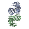
| ||||||||||||
|---|---|---|---|---|---|---|---|---|---|---|---|---|---|
| 1 | 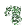
| ||||||||||||
| 2 | 
| ||||||||||||
| Unit cell |
| ||||||||||||
| Noncrystallographic symmetry (NCS) | NCS oper:
| ||||||||||||
| Details | MTRIX THE TRANSFORMATIONS PRESENTED ON MTRIX RECORDS BELOW DESCRIBE NON-CRYSTALLOGRAPHIC RELATIONSHIPS AMONG THE VARIOUS DOMAINS IN THIS ENTRY. APPLYING THE APPROPRIATE MTRIX TRANSFORMATION TO THE RESIDUES LISTED FIRST WILL YIELD APPROXIMATE COORDINATES FOR THE RESIDUES LISTED SECOND. APPLIED TO TRANSFORMED TO MTRIX RESIDUES RESIDUES RMSD M1 B 1 .. B 420 A 1 .. A 420 0.426 M2 B 421 .. B 561 A 421 .. A 561 1.117 M1 SUPERIMPOSES DOMAINS I - III OF MONOMER B ON THE CORRESPONDING DOMAINS OF MONOMER A, BASED ON A LEAST SQUARES PROCEDURE (HOMOLOGY, ROSSMANN AND ARGOS,1975) USING 301 SELECTED ALPHA-CARBON ATOMS IN THESE DOMAINS. M2 SUPERIMPOSES DOMAIN IV OF MONOMER B ON DOMAIN IV OF MONOMER A, USING 35 SELECTED ALPHA-CARBON ATOMS FROM THE CENTRAL PART OF THE BETA-SHEET IN THESE DOMAINS. |
-Components
| #1: Protein | Mass: 61579.902 Da / Num. of mol.: 2 / Source method: isolated from a natural source / Source: (natural) References: UniProt: P00949, phosphoglucomutase (alpha-D-glucose-1,6-bisphosphate-dependent) #2: Chemical |   Mass: 24.305 Da / Num. of mol.: 2 / Source method: obtained synthetically / Formula: Mg Mass: 24.305 Da / Num. of mol.: 2 / Source method: obtained synthetically / Formula: Mg#3: Water | ChemComp-HOH / |  Mass: 18.015 Da / Num. of mol.: 494 / Source method: isolated from a natural source / Formula: H2O Mass: 18.015 Da / Num. of mol.: 494 / Source method: isolated from a natural source / Formula: H2OHas protein modification | Y | |
|---|
-Experimental details
-Experiment
| Experiment | Method: X-RAY DIFFRACTION |
|---|
- Sample preparation
| Crystal | Density Matthews: 3.12 Å3/Da / Density % sol: 60.59 % | ||||||||||||||||||||||||
|---|---|---|---|---|---|---|---|---|---|---|---|---|---|---|---|---|---|---|---|---|---|---|---|---|---|
| Crystal grow | *PLUS pH: 6.5 / Method: unknown | ||||||||||||||||||||||||
| Components of the solutions | *PLUS
|
-Data collection
| Diffraction source | Wavelength: 1.5418 Å |
|---|---|
| Detector | Type: XUONG-HAMLIN MULTIWIRE / Detector: AREA DETECTOR / Date: Oct 1, 1993 |
| Radiation | Monochromatic (M) / Laue (L): M / Scattering type: x-ray |
| Radiation wavelength | Wavelength: 1.5418 Å / Relative weight: 1 |
| Reflection | Resolution: 2.4→13.1 Å / Num. obs: 54770 / % possible obs: 90 % / Observed criterion σ(I): 1 / Redundancy: 4.1 % / Rmerge(I) obs: 0.054 |
| Reflection | *PLUS Highest resolution: 2.35 Å / Lowest resolution: 20 Å / Num. obs: 54817 / % possible obs: 84.2 % / Rmerge(I) obs: 0.054 |
| Reflection shell | *PLUS Highest resolution: 2.35 Å / Lowest resolution: 2.41 Å / % possible obs: 56.8 % |
- Processing
| Software |
| ||||||||||||||||||||||||||||||||||||||||||||||||||||||||||||
|---|---|---|---|---|---|---|---|---|---|---|---|---|---|---|---|---|---|---|---|---|---|---|---|---|---|---|---|---|---|---|---|---|---|---|---|---|---|---|---|---|---|---|---|---|---|---|---|---|---|---|---|---|---|---|---|---|---|---|---|---|---|
| Refinement | Resolution: 2.4→6 Å / σ(F): 1 Details: THE MODEL CONTAINS FIVE RESIDUES OUT OF 1122 THAT FALL IN THE GENEROUSLY ALLOWED REGION OF A RAMACHANDRAN PLOT AS DEFINED IN PROCHECK AND ONE RESIDUE IN THE DISALLOWED REGION. TWO OF THE ...Details: THE MODEL CONTAINS FIVE RESIDUES OUT OF 1122 THAT FALL IN THE GENEROUSLY ALLOWED REGION OF A RAMACHANDRAN PLOT AS DEFINED IN PROCHECK AND ONE RESIDUE IN THE DISALLOWED REGION. TWO OF THE FIVE ARE ACTIVE SITE RESIDUES IN CHAIN A AND B AND THE OTHER FOUR ARE INVOLVED IN CRYSTALLOGRAPHIC CONTACTS. THE RESIDUES ARE ARG A 216, SER A 116, ASN A 461, SER B 116, AND ASN B 461.
| ||||||||||||||||||||||||||||||||||||||||||||||||||||||||||||
| Displacement parameters | Biso mean: 28 Å2 | ||||||||||||||||||||||||||||||||||||||||||||||||||||||||||||
| Refinement step | Cycle: LAST / Resolution: 2.4→6 Å
| ||||||||||||||||||||||||||||||||||||||||||||||||||||||||||||
| Refine LS restraints |
| ||||||||||||||||||||||||||||||||||||||||||||||||||||||||||||
| Software | *PLUS Name: X-PLOR / Classification: refinement | ||||||||||||||||||||||||||||||||||||||||||||||||||||||||||||
| Refinement | *PLUS | ||||||||||||||||||||||||||||||||||||||||||||||||||||||||||||
| Solvent computation | *PLUS | ||||||||||||||||||||||||||||||||||||||||||||||||||||||||||||
| Displacement parameters | *PLUS | ||||||||||||||||||||||||||||||||||||||||||||||||||||||||||||
| Refine LS restraints | *PLUS
|
