 Movie
Movie Controller

[English] 日本語
 Yorodumi
- PDB-4bub: CRYSTAL STRUCTURE OF MURE LIGASE FROM THERMOTOGA MARITIMA IN COMP... -
+ Open data

- Basic information
| Entry | Database: PDB / ID: 4bub | ||||||
|---|---|---|---|---|---|---|---|
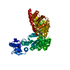
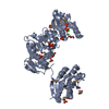
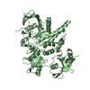
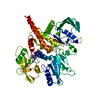
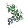
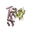
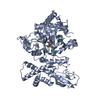
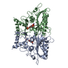
| Title | CRYSTAL STRUCTURE OF MURE LIGASE FROM THERMOTOGA MARITIMA IN COMPLEX WITH ADP | ||||||
 Components Components | UDP-N-ACETYLMURAMOYL-L-ALANYL-D-GLUTAMATE--LD-LYSINE LIGASE | ||||||
 Keywords Keywords | LIGASE / PEPTIDOGLYCAN SYNTHESIS / MURE / ADP-FORMING ENZYME / CELL WALL / CELL SHAPE / CELL CYCLE / NUCLEOTIDE-BINDING / ATP- BINDING / CELL DIVISION | ||||||
| Function / homology |  Function and homology information Function and homology informationUDP-N-acetylmuramoyl-L-alanyl-D-glutamate-D-lysine ligase / UDP-N-acetylmuramoyl-L-alanyl-D-glutamate--D-lysine ligase activity / UDP-N-acetylmuramoyl-L-alanyl-D-glutamate-L-lysine ligase / UDP-N-acetylmuramoyl-L-alanyl-D-glutamate-L-lysine ligase activity / tetrahydrofolylpolyglutamate synthase activity / peptidoglycan biosynthetic process / cell wall organization / regulation of cell shape / cell division / magnesium ion binding ...UDP-N-acetylmuramoyl-L-alanyl-D-glutamate-D-lysine ligase / UDP-N-acetylmuramoyl-L-alanyl-D-glutamate--D-lysine ligase activity / UDP-N-acetylmuramoyl-L-alanyl-D-glutamate-L-lysine ligase / UDP-N-acetylmuramoyl-L-alanyl-D-glutamate-L-lysine ligase activity / tetrahydrofolylpolyglutamate synthase activity / peptidoglycan biosynthetic process / cell wall organization / regulation of cell shape / cell division / magnesium ion binding / ATP binding / cytoplasm Similarity search - Function | ||||||
| Biological species |   THERMOTOGA MARITIMA (bacteria) THERMOTOGA MARITIMA (bacteria) | ||||||
| Method |  X-RAY DIFFRACTION / SYNCHROTRON / SAD / Resolution: 2.9 Å X-RAY DIFFRACTION / SYNCHROTRON / SAD / Resolution: 2.9 Å | ||||||
 Authors Authors | Favini-Stabile, S. / Contreras-Martel, C. / Thielens, N. / Dessen, A. | ||||||
 Citation Citation | Journal: Environ.Microbiol. / Year: 2013 Title: Mreb and Murg as Scaffolds for the Cytoplasmic Steps of Peptidoglycan Biosynthesis Authors: Favini-Stabile, S. / Contreras-Martel, C. / Thielens, N. / Dessen, A. | ||||||
| History |
|
- Structure visualization
| Structure viewer | Molecule: MolmilJmol/JSmol |
|---|
- Downloads & links
-Download
| PDBx/mmCIF format | 4bub.cif.gz | 202.8 KB | Display | PDBx/mmCIF format |
|---|---|---|---|---|
| PDB format | pdb4bub.ent.gz | 162.4 KB | Display | PDB format |
| PDBx/mmJSON format | 4bub.json.gz | Tree view | PDBx/mmJSON format | |
| Others |  Other downloads Other downloads |
-Validation report
| Arichive directory | https://data.pdbj.org/pub/pdb/validation_reports/bu/4bubftp://data.pdbj.org/pub/pdb/validation_reports/bu/4bub | HTTPS FTP |
|---|
-Related structure data


-Links
 PDBj
PDBj- Assembly
| Deposited unit | 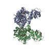
| |||||||||||||||||||||||||||||||||||||||||||||||||||||||||||||||||||||||||||||||||||||||||||||||
|---|---|---|---|---|---|---|---|---|---|---|---|---|---|---|---|---|---|---|---|---|---|---|---|---|---|---|---|---|---|---|---|---|---|---|---|---|---|---|---|---|---|---|---|---|---|---|---|---|---|---|---|---|---|---|---|---|---|---|---|---|---|---|---|---|---|---|---|---|---|---|---|---|---|---|---|---|---|---|---|---|---|---|---|---|---|---|---|---|---|---|---|---|---|---|---|---|
| 1 | 
| |||||||||||||||||||||||||||||||||||||||||||||||||||||||||||||||||||||||||||||||||||||||||||||||
| 2 | 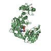
| |||||||||||||||||||||||||||||||||||||||||||||||||||||||||||||||||||||||||||||||||||||||||||||||
| Unit cell |
| |||||||||||||||||||||||||||||||||||||||||||||||||||||||||||||||||||||||||||||||||||||||||||||||
| Noncrystallographic symmetry (NCS) | NCS domain:
NCS domain segments:
NCS ensembles :
NCS oper:
|
-Components
| #1: Protein | Mass: 56309.523 Da / Num. of mol.: 2 Source method: isolated from a genetically manipulated source Source: (gene. exp.) THERMOTOGA MARITIMA (bacteria) / Strain: DSM 3109 / Production host: References: UniProt: Q9WY79, UDP-N-acetylmuramoyl-L-alanyl-D-glutamate-D-lysine ligase, UDP-N-acetylmuramoyl-L-alanyl-D-glutamate-L-lysine ligase #2: Chemical |   Mass: 427.201 Da / Num. of mol.: 2 / Source method: obtained synthetically / Formula: C10H15N5O10P2 / Comment: ADP, energy-carrying molecule*YM Mass: 427.201 Da / Num. of mol.: 2 / Source method: obtained synthetically / Formula: C10H15N5O10P2 / Comment: ADP, energy-carrying molecule*YM#3: Chemical |   Mass: 24.305 Da / Num. of mol.: 2 / Source method: obtained synthetically / Formula: Mg Mass: 24.305 Da / Num. of mol.: 2 / Source method: obtained synthetically / Formula: Mg#4: Water | ChemComp-HOH / |  Mass: 18.015 Da / Num. of mol.: 17 / Source method: isolated from a natural source / Formula: H2O Mass: 18.015 Da / Num. of mol.: 17 / Source method: isolated from a natural source / Formula: H2OHas protein modification | Y | |
|---|
-Experimental details
-Experiment
| Experiment | Method: X-RAY DIFFRACTION / Number of used crystals: 1 |
|---|
- Sample preparation
| Crystal | Density Matthews: 3.24 Å3/Da / Density % sol: 62 % / Description: NONE |
|---|---|
| Crystal grow | pH: 5 / Details: 0.1M CITIRC ACID PH5, 3% PEGMME 5000 |
-Data collection
| Diffraction | Mean temperature: 100 K |
|---|---|
| Diffraction source | Source: SYNCHROTRON / Site: ESRF  / Beamline: ID14-4 / Wavelength: 0.97921 / Beamline: ID14-4 / Wavelength: 0.97921 |
| Detector | Type: ADSC QUANTUM 315r / Detector: CCD / Date: Nov 17, 2012 |
| Radiation | Protocol: SINGLE WAVELENGTH / Monochromatic (M) / Laue (L): M / Scattering type: x-ray |
| Radiation wavelength | Wavelength: 0.97921 Å / Relative weight: 1 |
| Reflection | Resolution: 2.9→20 Å / Num. obs: 29228 / % possible obs: 95.9 % / Observed criterion σ(I): 3 / Redundancy: 11 % / Biso Wilson estimate: 94.853 Å2 / Rmerge(I) obs: 0.06 / Net I/σ(I): 40 |
| Reflection shell | Resolution: 2.9→3.07 Å / Redundancy: 12.1 % / Rmerge(I) obs: 0.1 / Mean I/σ(I) obs: 3 / % possible all: 81 |
- Processing
| Software |
| ||||||||||||||||||||||||||||||||||||||||||||||||||||||||||||||||||||||||||||||||||||||||||||||||||||||||||||||||||||||||||||||||||||||||||||||||||||||||||||||||||||||||||||||||||||||
|---|---|---|---|---|---|---|---|---|---|---|---|---|---|---|---|---|---|---|---|---|---|---|---|---|---|---|---|---|---|---|---|---|---|---|---|---|---|---|---|---|---|---|---|---|---|---|---|---|---|---|---|---|---|---|---|---|---|---|---|---|---|---|---|---|---|---|---|---|---|---|---|---|---|---|---|---|---|---|---|---|---|---|---|---|---|---|---|---|---|---|---|---|---|---|---|---|---|---|---|---|---|---|---|---|---|---|---|---|---|---|---|---|---|---|---|---|---|---|---|---|---|---|---|---|---|---|---|---|---|---|---|---|---|---|---|---|---|---|---|---|---|---|---|---|---|---|---|---|---|---|---|---|---|---|---|---|---|---|---|---|---|---|---|---|---|---|---|---|---|---|---|---|---|---|---|---|---|---|---|---|---|---|---|
| Refinement | Method to determine structure: SAD Starting model: NONE Resolution: 2.9→48.48 Å / Cor.coef. Fo:Fc: 0.955 / Cor.coef. Fo:Fc free: 0.919 / SU B: 25.587 / SU ML: 0.445 / Cross valid method: THROUGHOUT / ESU R Free: 0.461 / Stereochemistry target values: MAXIMUM LIKELIHOOD / Details: HYDROGENS HAVE BEEN ADDED IN THE RIDING POSITIONS.
| ||||||||||||||||||||||||||||||||||||||||||||||||||||||||||||||||||||||||||||||||||||||||||||||||||||||||||||||||||||||||||||||||||||||||||||||||||||||||||||||||||||||||||||||||||||||
| Solvent computation | Ion probe radii: 0.8 Å / Shrinkage radii: 0.8 Å / VDW probe radii: 1.2 Å / Solvent model: BABINET MODEL WITH MASK | ||||||||||||||||||||||||||||||||||||||||||||||||||||||||||||||||||||||||||||||||||||||||||||||||||||||||||||||||||||||||||||||||||||||||||||||||||||||||||||||||||||||||||||||||||||||
| Displacement parameters | Biso mean: 110.399 Å2
| ||||||||||||||||||||||||||||||||||||||||||||||||||||||||||||||||||||||||||||||||||||||||||||||||||||||||||||||||||||||||||||||||||||||||||||||||||||||||||||||||||||||||||||||||||||||
| Refinement step | Cycle: LAST / Resolution: 2.9→48.48 Å
| ||||||||||||||||||||||||||||||||||||||||||||||||||||||||||||||||||||||||||||||||||||||||||||||||||||||||||||||||||||||||||||||||||||||||||||||||||||||||||||||||||||||||||||||||||||||
| Refine LS restraints |
|