 Movie
Movie Controller


+ Open data

- Basic information
| Entry | Database: PDB / ID: 2wk1 | ||||||
|---|---|---|---|---|---|---|---|
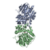
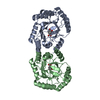
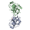
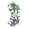
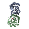
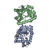
| Title | Structure of the O-methyltransferase NovP | ||||||
 Components Components | NOVP | ||||||
 Keywords Keywords | TRANSFERASE / NOVP / O-METHYLTRANSFERASE / NOVOBIOCIN / TYLF SUPERFAMILY | ||||||
| Function / homology |  Function and homology information Function and homology informationdemethyldecarbamoylnovobiocin O-methyltransferase / novobiocin biosynthetic process / methyltransferase activity / methylation / metal ion binding Similarity search - Function | ||||||
| Biological species |  STREPTOMYCES CAERULEUS (bacteria) STREPTOMYCES CAERULEUS (bacteria) | ||||||
| Method |  X-RAY DIFFRACTION / SYNCHROTRON / MOLECULAR REPLACEMENT / Resolution: 1.4 Å X-RAY DIFFRACTION / SYNCHROTRON / MOLECULAR REPLACEMENT / Resolution: 1.4 Å | ||||||
 Authors Authors | Gomez Garcia, I. / Stevenson, C.E.M. / Uson, I. / Freel Meyers, C.L. / Walsh, C.T. / Lawson, D.M. | ||||||
 Citation Citation | Journal: J.Mol.Biol. / Year: 2010 Title: The Crystal Structure of the Novobiocin Biosynthetic Enzyme Novp: The First Representative Structure for the Tylf O-Methyltransferase Superfamily. Authors: Gomez Garcia, I. / Stevenson, C.E.M. / Uson, I. / Freel Meyers, C.L. / Walsh, C.T. / Lawson, D.M. #1: Journal: Acta Crystallogr.,Sect.F / Year: 2007 Title: Crystallization and Preliminary X-Ray Analysis of the O-Methyltransferase Novp from the Novobiocin-Biosynthetic Cluster of Streptomyces Spheroides. Authors: Stevenson, C.E.M. / Freel Meyers, C.L. / Walsh, C.T. / Lawson, D.M. #2: Journal: Acta Crystallogr.,Sect.D / Year: 2007 Title: Structure Determination of the O-Methyltransferase Novp Using the 'Free Lunch Algorithm' as Implemented in Shelxe. Authors: Uson, I. / Stevenson, C.E.M. / Lawson, D.M. / Sheldrick, G.M. | ||||||
| History |
|
- Structure visualization
| Structure viewer | Molecule: MolmilJmol/JSmol |
|---|
- Downloads & links
-Download
| PDBx/mmCIF format | 2wk1.cif.gz | 124.7 KB | Display | PDBx/mmCIF format |
|---|---|---|---|---|
| PDB format | pdb2wk1.ent.gz | 95.4 KB | Display | PDB format |
| PDBx/mmJSON format | 2wk1.json.gz | Tree view | PDBx/mmJSON format | |
| Others |  Other downloads Other downloads |
-Validation report
| Arichive directory | https://data.pdbj.org/pub/pdb/validation_reports/wk/2wk1ftp://data.pdbj.org/pub/pdb/validation_reports/wk/2wk1 | HTTPS FTP |
|---|
-Related structure data
| Related structure data |  1vidS S: Starting model for refinement |
|---|---|
| Similar structure data |


-Links
 PDBj
PDBj- Assembly
| Deposited unit | 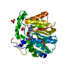
| ||||||||
|---|---|---|---|---|---|---|---|---|---|
| 1 | 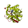
| ||||||||
| Unit cell |
| ||||||||
| Components on special symmetry positions |
|
-Components
| #1: Protein | Mass: 32175.088 Da / Num. of mol.: 1 Source method: isolated from a genetically manipulated source Details: DISULPHIDE BRIDGE BETWEEN CYS228 AND CYS231 / Source: (gene. exp.) STREPTOMYCES CAERULEUS (bacteria) / Production host: References: UniProt: Q9L9F2, Transferases; Transferring one-carbon groups; Methyltransferases | ||||||
|---|---|---|---|---|---|---|---|
| #2: Chemical | ChemComp-SAH /   Mass: 384.411 Da / Num. of mol.: 1 / Source method: obtained synthetically / Formula: C14H20N6O5S Mass: 384.411 Da / Num. of mol.: 1 / Source method: obtained synthetically / Formula: C14H20N6O5S | ||||||
| #3: Chemical |   Mass: 96.063 Da / Num. of mol.: 2 / Source method: obtained synthetically / Formula: SO4 Mass: 96.063 Da / Num. of mol.: 2 / Source method: obtained synthetically / Formula: SO4#4: Chemical |   Mass: 62.068 Da / Num. of mol.: 2 / Source method: obtained synthetically / Formula: C2H6O2 Mass: 62.068 Da / Num. of mol.: 2 / Source method: obtained synthetically / Formula: C2H6O2#5: Water | ChemComp-HOH / |  Mass: 18.015 Da / Num. of mol.: 242 / Source method: isolated from a natural source / Formula: H2O Mass: 18.015 Da / Num. of mol.: 242 / Source method: isolated from a natural source / Formula: H2OHas protein modification | Y | |
-Experimental details
-Experiment
| Experiment | Method: X-RAY DIFFRACTION / Number of used crystals: 1 |
|---|
- Sample preparation
| Crystal | Density Matthews: 2.19 Å3/Da / Density % sol: 43.9 % / Description: NONE |
|---|---|
| Crystal grow | pH: 7 / Details: 2 M AMMONIUM SULPHATE, 100 MM HEPES BUFFER PH 7.0 |
-Data collection
| Diffraction | Mean temperature: 100 K |
|---|---|
| Diffraction source | Source: SYNCHROTRON / Site: SRS  / Beamline: PX10.1 / Wavelength: 1.488 / Beamline: PX10.1 / Wavelength: 1.488 |
| Detector | Type: MARRESEARCH / Detector: CCD / Date: Apr 14, 2005 |
| Radiation | Protocol: SINGLE WAVELENGTH / Monochromatic (M) / Laue (L): M / Scattering type: x-ray |
| Radiation wavelength | Wavelength: 1.488 Å / Relative weight: 1 |
| Reflection | Resolution: 1.4→36.3 Å / Num. obs: 54638 / % possible obs: 98.8 % / Observed criterion σ(I): -3 / Redundancy: 3.4 % / Biso Wilson estimate: 17.6 Å2 / Rmerge(I) obs: 0.06 / Net I/σ(I): 18.1 |
| Reflection shell | Resolution: 1.4→1.42 Å / Rmerge(I) obs: 0.16 / Mean I/σ(I) obs: 4.2 / % possible all: 89.5 |
- Processing
| Software |
| ||||||||||||||||||||||||||||||||||||||||||||||||||||||||||||||||||||||||||||||||||||||||||||||||||||||||||||||||||||||||||||||||||||||||||||||||||||||||||||||||||||||||||||||||||||||
|---|---|---|---|---|---|---|---|---|---|---|---|---|---|---|---|---|---|---|---|---|---|---|---|---|---|---|---|---|---|---|---|---|---|---|---|---|---|---|---|---|---|---|---|---|---|---|---|---|---|---|---|---|---|---|---|---|---|---|---|---|---|---|---|---|---|---|---|---|---|---|---|---|---|---|---|---|---|---|---|---|---|---|---|---|---|---|---|---|---|---|---|---|---|---|---|---|---|---|---|---|---|---|---|---|---|---|---|---|---|---|---|---|---|---|---|---|---|---|---|---|---|---|---|---|---|---|---|---|---|---|---|---|---|---|---|---|---|---|---|---|---|---|---|---|---|---|---|---|---|---|---|---|---|---|---|---|---|---|---|---|---|---|---|---|---|---|---|---|---|---|---|---|---|---|---|---|---|---|---|---|---|---|---|
| Refinement | Method to determine structure: MOLECULAR REPLACEMENT Starting model: PDB ENTRY 1VID Resolution: 1.4→34.1 Å / Cor.coef. Fo:Fc: 0.975 / Cor.coef. Fo:Fc free: 0.973 / SU B: 1.321 / SU ML: 0.024 / Cross valid method: THROUGHOUT / σ(F): 0 / ESU R: 0.055 / ESU R Free: 0.048 / Stereochemistry target values: MAXIMUM LIKELIHOOD Details: HYDROGENS HAVE BEEN ADDED IN THE RIDING POSITIONS. DATA WERE COLLECTED TO A MAXIMUM RESOLUTION OF 1.35 ANGSTROM, BUT WERE NOT VERY COMPLETE IN THE OUTER RESOLUTION SHELL, SO ONLY THE DATA TO ...Details: HYDROGENS HAVE BEEN ADDED IN THE RIDING POSITIONS. DATA WERE COLLECTED TO A MAXIMUM RESOLUTION OF 1.35 ANGSTROM, BUT WERE NOT VERY COMPLETE IN THE OUTER RESOLUTION SHELL, SO ONLY THE DATA TO 1.4 ANGSTROM RESOLUTION WERE USED FOR THE REFINEMENT. HOWEVER, THE STRUCTURE WAS SOLVED USING ALL AVAILABLE NATIVE DATA INCLUDING THREE OTHER NATIVE DATA SETS COLLECTED AT LOWER RESOLUTIONS. THIS MERGED NATIVE DATA SET HAS ALSO BEEN DEPOSITED. SEE REFERENCE 2 (USON ET AL., 2007) FOR FURTHER DETAILS.
| ||||||||||||||||||||||||||||||||||||||||||||||||||||||||||||||||||||||||||||||||||||||||||||||||||||||||||||||||||||||||||||||||||||||||||||||||||||||||||||||||||||||||||||||||||||||
| Solvent computation | Ion probe radii: 0.8 Å / VDW probe radii: 1.4 Å / Solvent model: MASK BULK SOLVENT | ||||||||||||||||||||||||||||||||||||||||||||||||||||||||||||||||||||||||||||||||||||||||||||||||||||||||||||||||||||||||||||||||||||||||||||||||||||||||||||||||||||||||||||||||||||||
| Displacement parameters | Biso mean: 13.38 Å2
| ||||||||||||||||||||||||||||||||||||||||||||||||||||||||||||||||||||||||||||||||||||||||||||||||||||||||||||||||||||||||||||||||||||||||||||||||||||||||||||||||||||||||||||||||||||||
| Refinement step | Cycle: LAST / Resolution: 1.4→34.1 Å
| ||||||||||||||||||||||||||||||||||||||||||||||||||||||||||||||||||||||||||||||||||||||||||||||||||||||||||||||||||||||||||||||||||||||||||||||||||||||||||||||||||||||||||||||||||||||
| Refine LS restraints |
|