 Movie
Movie Controller

[English] 日本語
 Yorodumi
- PDB-4bow: Crystal structure of LamA_E269S from Z. galactanivorans in comple... -
+ Open data

- Basic information
| Entry | Database: PDB / ID: 4bow | |||||||||
|---|---|---|---|---|---|---|---|---|---|---|
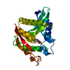
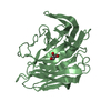
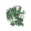
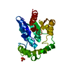
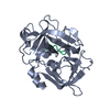
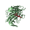
| Title | Crystal structure of LamA_E269S from Z. galactanivorans in complex with laminaritriose and laminaritetraose | |||||||||
 Components Components | ENDO-1,3-BETA-GLUCANASE, FAMILY GH16 | |||||||||
 Keywords Keywords | HYDROLASE / MARINE LAMINARINASE / FAMILY GH16 GLYCOSIDE HYDROLASE | |||||||||
| Function / homology |  Function and homology information Function and homology informationglucan endo-1,3-beta-D-glucosidase / glucan endo-1,3-beta-D-glucosidase activity / carbohydrate metabolic process / metal ion binding Similarity search - Function | |||||||||
| Biological species |  ZOBELLIA GALACTANIVORANS (bacteria) ZOBELLIA GALACTANIVORANS (bacteria) | |||||||||
| Method |  X-RAY DIFFRACTION / SYNCHROTRON / MOLECULAR REPLACEMENT / Resolution: 1.35 Å X-RAY DIFFRACTION / SYNCHROTRON / MOLECULAR REPLACEMENT / Resolution: 1.35 Å | |||||||||
 Authors Authors | Labourel, A. / Jeudy, A. / Czjzek, M. / Michel, G. | |||||||||
 Citation Citation | Journal: J.Biol.Chem. / Year: 2014 Title: The Beta-Glucanase Zglama from Zobellia Galactanivorans Evolved a Bent Active Site Adapted for Efficient Degradation of Algal Laminarin Authors: Labourel, A. / Jam, M. / Jeudy, A. / Hehemann, J.H. / Czjzek, M. / Michel, G. | |||||||||
| History |
|
- Structure visualization
| Structure viewer | Molecule: MolmilJmol/JSmol |
|---|
- Downloads & links
-Download
| PDBx/mmCIF format | 4bow.cif.gz | 247.2 KB | Display | PDBx/mmCIF format |
|---|---|---|---|---|
| PDB format | pdb4bow.ent.gz | 197.1 KB | Display | PDB format |
| PDBx/mmJSON format | 4bow.json.gz | Tree view | PDBx/mmJSON format | |
| Others |  Other downloads Other downloads |
-Validation report
| Arichive directory | https://data.pdbj.org/pub/pdb/validation_reports/bo/4bowftp://data.pdbj.org/pub/pdb/validation_reports/bo/4bow | HTTPS FTP |
|---|
-Related structure data
| Related structure data | 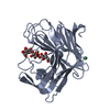 4bpzC 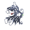 4bq1SC C: citing same article ( S: Starting model for refinement |
|---|---|
| Similar structure data |


-Links
 PDBj
PDBj


- Assembly
| Deposited unit | 
| ||||||||||||||||
|---|---|---|---|---|---|---|---|---|---|---|---|---|---|---|---|---|---|
| 1 | 
| ||||||||||||||||
| 2 | 
| ||||||||||||||||
| Unit cell |
| ||||||||||||||||
| Noncrystallographic symmetry (NCS) | NCS oper:
|
-Components
-Protein , 1 types, 2 molecules AB
| #1: Protein | Mass: 29219.875 Da / Num. of mol.: 2 / Fragment: CATALYTIC DOMAIN, RESIDUES 136-383 / Mutation: YES Source method: isolated from a genetically manipulated source Source: (gene. exp.) ZOBELLIA GALACTANIVORANS (bacteria) / Strain: DSIJTDescription: GERMAN COLLECTION OF MICROORGANISMS (DSM) AND IN HOUSE AT ROSCOFF COLLECTION Production host: References: UniProt: G0L5X4, glucan endo-1,3-beta-D-glucosidase |
|---|
-Sugars , 3 types, 3 molecules 
| #2: Polysaccharide | beta-D-glucopyranose-(1-3)-beta-D-glucopyranose-(1-3)-alpha-D-glucopyranose Source method: isolated from a genetically manipulated source |
|---|---|
| #3: Polysaccharide | beta-D-glucopyranose-(1-3)-beta-D-glucopyranose-(1-3)-beta-D-glucopyranose-(1-3)-beta-D-glucopyranose Source method: isolated from a genetically manipulated source |
| #6: Sugar | ChemComp-BGC /  Type: D-saccharide, beta linking / Mass: 180.156 Da / Num. of mol.: 1 Type: D-saccharide, beta linking / Mass: 180.156 Da / Num. of mol.: 1Source method: isolated from a genetically manipulated source Formula: C6H12O6 |
-Non-polymers , 3 types, 548 molecules 


| #4: Chemical |  Mass: 40.078 Da / Num. of mol.: 2 / Source method: obtained synthetically / Formula: Ca Mass: 40.078 Da / Num. of mol.: 2 / Source method: obtained synthetically / Formula: Ca#5: Chemical | ChemComp-NA / |  Mass: 22.990 Da / Num. of mol.: 1 / Source method: obtained synthetically / Formula: Na Mass: 22.990 Da / Num. of mol.: 1 / Source method: obtained synthetically / Formula: Na#7: Water | ChemComp-HOH / | Mass: 18.015 Da / Num. of mol.: 545 / Source method: isolated from a natural source / Formula: H2O |
|---|
-Details
| Sequence details | THE CONSTRUCTION CONTAINS THE CATALYTIC MODULE ONLY. THE N- TERMINAL PKD MODULE HAS NOT BEEN CLONED. ...THE CONSTRUCTI |
|---|
-Experimental details
-Experiment
| Experiment | Method: X-RAY DIFFRACTION / Number of used crystals: 1 |
|---|
- Sample preparation
| Crystal | Density Matthews: 2.08 Å3/Da / Density % sol: 52 % / Description: NONE |
|---|---|
| Crystal grow | Temperature: 293 K / Method: vapor diffusion, hanging drop / pH: 4 Details: 100 MM MIB PH 4.0, 19% PEG1500, HANGING DROP AT 20 DEGREES CELSIUS MIXING 2 MICROL OF PROTEIN (13.7 MG/ML SUPPLEMENTED WITH 5 MM PURIFIED LAMINARIHEXAOSE) WITH 1 MICROL OF RESERVOIR SOLUTION |
-Data collection
| Diffraction | Mean temperature: 100 K |
|---|---|
| Diffraction source | Source: SYNCHROTRON / Site: ESRF  / Beamline: ID29 / Wavelength: 0.97625 / Beamline: ID29 / Wavelength: 0.97625 |
| Detector | Type: DECTRIS PILATUS 6M / Detector: PIXEL / Date: Jul 7, 2011 |
| Radiation | Protocol: SINGLE WAVELENGTH / Monochromatic (M) / Laue (L): M / Scattering type: x-ray |
| Radiation wavelength | Wavelength: 0.97625 Å / Relative weight: 1 |
| Reflection | Resolution: 1.35→40.44 Å / Num. obs: 107451 / % possible obs: 99.7 % / Observed criterion σ(I): 0 / Redundancy: 4.7 % / Rmerge(I) obs: 0.04 / Net I/σ(I): 16.3 |
| Reflection shell | Resolution: 1.35→1.42 Å / Redundancy: 4.7 % / Rmerge(I) obs: 0.55 / Mean I/σ(I) obs: 2.3 / % possible all: 99.7 |
- Processing
| Software |
| ||||||||||||||||||||||||||||||||||||||||||||||||||||||||||||||||||||||||||||||||||||||||||||||||||||||||||||||||||||||||||||||||||||||||||||||||||||||||||||||||||||||||||||||||||||||
|---|---|---|---|---|---|---|---|---|---|---|---|---|---|---|---|---|---|---|---|---|---|---|---|---|---|---|---|---|---|---|---|---|---|---|---|---|---|---|---|---|---|---|---|---|---|---|---|---|---|---|---|---|---|---|---|---|---|---|---|---|---|---|---|---|---|---|---|---|---|---|---|---|---|---|---|---|---|---|---|---|---|---|---|---|---|---|---|---|---|---|---|---|---|---|---|---|---|---|---|---|---|---|---|---|---|---|---|---|---|---|---|---|---|---|---|---|---|---|---|---|---|---|---|---|---|---|---|---|---|---|---|---|---|---|---|---|---|---|---|---|---|---|---|---|---|---|---|---|---|---|---|---|---|---|---|---|---|---|---|---|---|---|---|---|---|---|---|---|---|---|---|---|---|---|---|---|---|---|---|---|---|---|---|
| Refinement | Method to determine structure: MOLECULAR REPLACEMENT Starting model: PDB ENTRY 4BQ1 Resolution: 1.35→67.44 Å / Cor.coef. Fo:Fc: 0.982 / Cor.coef. Fo:Fc free: 0.974 / SU B: 1.88 / SU ML: 0.034 / Cross valid method: THROUGHOUT / ESU R: 0.051 / ESU R Free: 0.049 / Stereochemistry target values: MAXIMUM LIKELIHOOD Details: HYDROGENS HAVE BEEN ADDED IN THE RIDING POSITIONS. HYDROGENS HAVE BEEN USED IF PRESENT IN THE INPUT U VALUES, WITH TLS ADDED
| ||||||||||||||||||||||||||||||||||||||||||||||||||||||||||||||||||||||||||||||||||||||||||||||||||||||||||||||||||||||||||||||||||||||||||||||||||||||||||||||||||||||||||||||||||||||
| Solvent computation | Ion probe radii: 0.8 Å / Shrinkage radii: 0.8 Å / VDW probe radii: 1.2 Å / Solvent model: BABINET MODEL WITH MASK | ||||||||||||||||||||||||||||||||||||||||||||||||||||||||||||||||||||||||||||||||||||||||||||||||||||||||||||||||||||||||||||||||||||||||||||||||||||||||||||||||||||||||||||||||||||||
| Displacement parameters | Biso mean: 23.543 Å2
| ||||||||||||||||||||||||||||||||||||||||||||||||||||||||||||||||||||||||||||||||||||||||||||||||||||||||||||||||||||||||||||||||||||||||||||||||||||||||||||||||||||||||||||||||||||||
| Refinement step | Cycle: LAST / Resolution: 1.35→67.44 Å
| ||||||||||||||||||||||||||||||||||||||||||||||||||||||||||||||||||||||||||||||||||||||||||||||||||||||||||||||||||||||||||||||||||||||||||||||||||||||||||||||||||||||||||||||||||||||
| Refine LS restraints |
|