 Movie
Movie Controller

[English] 日本語
 Yorodumi
- PDB-2wfq: Crystal structure of the N-terminal signalling domain of human Dh... -
+ Open data

- Basic information
| Entry | Database: PDB / ID: 2wfq | ||||||
|---|---|---|---|---|---|---|---|
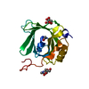
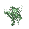
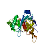
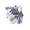
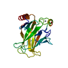
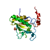
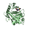
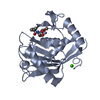
| Title | Crystal structure of the N-terminal signalling domain of human Dhh without calcium | ||||||
 Components Components | DESERT HEDGEHOG PROTEIN N-PRODUCT | ||||||
 Keywords Keywords | SIGNALING PROTEIN / LIPOPROTEIN / DEVELOPMENT / CELL MEMBRANE / AUTOCATALYTIC CLEAVAGE / DISEASE MUTATION / HEDGEHOG SIGNALLING / PROTEASE / MEMBRANE / SECRETED / PALMITATE / HYDROLASE / SIGNAL TRANSDUCTION / DEVELOPMENTAL PROTEIN | ||||||
| Function / homology |  Function and homology information Function and homology informationregulation of steroid biosynthetic process / cholesterol-protein transferase activity / HHAT G278V doesn't palmitoylate Hh-Np / Ligand-receptor interactions / Transcriptional regulation of testis differentiation / Leydig cell differentiation / male sex determination / patched binding / Activation of SMO / self proteolysis ...regulation of steroid biosynthetic process / cholesterol-protein transferase activity / HHAT G278V doesn't palmitoylate Hh-Np / Ligand-receptor interactions / Transcriptional regulation of testis differentiation / Leydig cell differentiation / male sex determination / patched binding / Activation of SMO / self proteolysis / Release of Hh-Np from the secreting cell / positive regulation of smoothened signaling pathway / cell fate specification / Class B/2 (Secretin family receptors) / smoothened signaling pathway / spermatid development / protein autoprocessing / myelination / Hedgehog ligand biogenesis / Hedgehog 'on' state / response to estrogen / osteoblast differentiation / response to estradiol / peptidase activity / cell-cell signaling / regulation of gene expression / Hydrolases; Acting on ester bonds / Golgi membrane / calcium ion binding / endoplasmic reticulum membrane / : / zinc ion binding / plasma membrane Similarity search - Function | ||||||
| Biological species |  HOMO SAPIENS (human) HOMO SAPIENS (human) | ||||||
| Method |  X-RAY DIFFRACTION / SYNCHROTRON / MOLECULAR REPLACEMENT / Resolution: 1.85 Å X-RAY DIFFRACTION / SYNCHROTRON / MOLECULAR REPLACEMENT / Resolution: 1.85 Å | ||||||
 Authors Authors | Bishop, B. / Aricescu, A.R. / Harlos, K. / O'Callaghan, C.A. / Jones, E.Y. / Siebold, C. | ||||||
 Citation Citation | Journal: Nat.Struct.Mol.Biol. / Year: 2009 Title: Structural Insights Into Hedgehog Ligand Sequestration by the Human Hedgehog-Interacting Protein Hip Authors: Bishop, B. / Aricescu, A.R. / Harlos, K. / O'Callaghan, C.A. / Jones, E.Y. / Siebold, C. | ||||||
| History |
|
- Structure visualization
| Structure viewer | Molecule: MolmilJmol/JSmol |
|---|
- Downloads & links
-Download
| PDBx/mmCIF format | 2wfq.cif.gz | 50 KB | Display | PDBx/mmCIF format |
|---|---|---|---|---|
| PDB format | pdb2wfq.ent.gz | 34 KB | Display | PDB format |
| PDBx/mmJSON format | 2wfq.json.gz | Tree view | PDBx/mmJSON format | |
| Others |  Other downloads Other downloads |
-Validation report
| Arichive directory | https://data.pdbj.org/pub/pdb/validation_reports/wf/2wfqftp://data.pdbj.org/pub/pdb/validation_reports/wf/2wfq | HTTPS FTP |
|---|
-Related structure data
| Related structure data |  2wfrC  2wftC  2wfxC  2wg3C  2wg4C  1vhhS C: citing same article ( S: Starting model for refinement |
|---|---|
| Similar structure data |


-Links
 PDBj
PDBj


- Assembly
| Deposited unit | 
| ||||||||
|---|---|---|---|---|---|---|---|---|---|
| 1 |
| ||||||||
| Unit cell |
|
-Components
| #1: Protein | Mass: 18849.082 Da / Num. of mol.: 1 / Fragment: N-TERMINAL SIGNALLING DOMAIN, RESIDUES 39-194 Source method: isolated from a genetically manipulated source Source: (gene. exp.) HOMO SAPIENS (human) / Plasmid: PET22B / Production host:  |
|---|---|
| #2: Chemical | ChemComp-ZN /   Mass: 65.409 Da / Num. of mol.: 1 / Source method: obtained synthetically / Formula: Zn Mass: 65.409 Da / Num. of mol.: 1 / Source method: obtained synthetically / Formula: Zn |
| #3: Chemical | ChemComp-SO4 /   Mass: 96.063 Da / Num. of mol.: 1 / Source method: obtained synthetically / Formula: SO4 Mass: 96.063 Da / Num. of mol.: 1 / Source method: obtained synthetically / Formula: SO4 |
| #4: Water | ChemComp-HOH /  Mass: 18.015 Da / Num. of mol.: 104 / Source method: isolated from a natural source / Formula: H2O Mass: 18.015 Da / Num. of mol.: 104 / Source method: isolated from a natural source / Formula: H2O |
-Experimental details
-Experiment
| Experiment | Method: X-RAY DIFFRACTION / Number of used crystals: 1 |
|---|
- Sample preparation
| Crystal | Density Matthews: 1.8 Å3/Da / Density % sol: 35.5 % / Description: NONE |
|---|---|
| Crystal grow | Details: 0.1 M TRIS-HCL, PH 8.5 0.2 M LITHIUM SULFATE 0.2 M SODIUM THIOCYANATE 30% (W/V) PEG4000 |
-Data collection
| Diffraction | Mean temperature: 100 K |
|---|---|
| Diffraction source | Source: SYNCHROTRON / Site: Diamond  / Beamline: I02 / Wavelength: 0.9507 / Beamline: I02 / Wavelength: 0.9507 |
| Detector | Type: ADSC CCD / Detector: CCD |
| Radiation | Protocol: SINGLE WAVELENGTH / Monochromatic (M) / Laue (L): M / Scattering type: x-ray |
| Radiation wavelength | Wavelength: 0.9507 Å / Relative weight: 1 |
| Reflection | Resolution: 1.85→30 Å / Num. obs: 12895 / % possible obs: 99.6 % / Observed criterion σ(I): 0 / Redundancy: 12.7 % / Rmerge(I) obs: 0.12 / Net I/σ(I): 14.6 |
| Reflection shell | Resolution: 1.85→1.95 Å / Redundancy: 12 % / Rmerge(I) obs: 0.68 / Mean I/σ(I) obs: 4.5 / % possible all: 99.7 |
- Processing
| Software |
| ||||||||||||||||||||||||||||||||||||||||||||||||||||||||||||||||||||||||||||||||||||||||||||||||||||||||||||||||||||||||||||||||||||||||||||||||||||||||||||||||||||||||||||||||||||||
|---|---|---|---|---|---|---|---|---|---|---|---|---|---|---|---|---|---|---|---|---|---|---|---|---|---|---|---|---|---|---|---|---|---|---|---|---|---|---|---|---|---|---|---|---|---|---|---|---|---|---|---|---|---|---|---|---|---|---|---|---|---|---|---|---|---|---|---|---|---|---|---|---|---|---|---|---|---|---|---|---|---|---|---|---|---|---|---|---|---|---|---|---|---|---|---|---|---|---|---|---|---|---|---|---|---|---|---|---|---|---|---|---|---|---|---|---|---|---|---|---|---|---|---|---|---|---|---|---|---|---|---|---|---|---|---|---|---|---|---|---|---|---|---|---|---|---|---|---|---|---|---|---|---|---|---|---|---|---|---|---|---|---|---|---|---|---|---|---|---|---|---|---|---|---|---|---|---|---|---|---|---|---|---|
| Refinement | Method to determine structure: MOLECULAR REPLACEMENT Starting model: PDB ENTRY 1VHH Resolution: 1.85→30 Å / Cor.coef. Fo:Fc: 0.957 / Cor.coef. Fo:Fc free: 0.931 / SU B: 7.983 / SU ML: 0.11 / TLS residual ADP flag: LIKELY RESIDUAL / Cross valid method: THROUGHOUT / ESU R: 0.165 / ESU R Free: 0.148 / Stereochemistry target values: MAXIMUM LIKELIHOOD / Details: HYDROGENS HAVE BEEN ADDED IN THE RIDING POSITIONS.
| ||||||||||||||||||||||||||||||||||||||||||||||||||||||||||||||||||||||||||||||||||||||||||||||||||||||||||||||||||||||||||||||||||||||||||||||||||||||||||||||||||||||||||||||||||||||
| Solvent computation | Ion probe radii: 0.8 Å / Shrinkage radii: 0.8 Å / VDW probe radii: 1.2 Å / Solvent model: MASK | ||||||||||||||||||||||||||||||||||||||||||||||||||||||||||||||||||||||||||||||||||||||||||||||||||||||||||||||||||||||||||||||||||||||||||||||||||||||||||||||||||||||||||||||||||||||
| Displacement parameters | Biso mean: 13.966 Å2
| ||||||||||||||||||||||||||||||||||||||||||||||||||||||||||||||||||||||||||||||||||||||||||||||||||||||||||||||||||||||||||||||||||||||||||||||||||||||||||||||||||||||||||||||||||||||
| Refinement step | Cycle: LAST / Resolution: 1.85→30 Å
| ||||||||||||||||||||||||||||||||||||||||||||||||||||||||||||||||||||||||||||||||||||||||||||||||||||||||||||||||||||||||||||||||||||||||||||||||||||||||||||||||||||||||||||||||||||||
| Refine LS restraints |
|