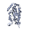
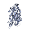
[English] 日本語
Yorodumi
- PDB-6kfa: Hydroxynitrile lyase from the millipede, Chamberlinius hualienens... -

+
Open data


ID or keywords:

Loading...

-
Basic information

Entry
Database: PDB / ID: 6kfa
TitleHydroxynitrile lyase from the millipede, Chamberlinius hualienensis bound with acetate
ComponentsHydroxynitrile lyase
KeywordsLYASE / Hydroxynitrile lyase / lipocalin fold / four disulfide bonds / dimer
Function / homologyRicin B-like lectins / lyase activity / ACETATE ION / Hydroxynitrile lyase
Function and homology information
Biological speciesChamberlinius hualienensis (arthropod)
MethodX-RAY DIFFRACTION / SYNCHROTRON / SAD / Resolution: 1.5 Å
AuthorsMotojima, F. / Izumi, A. / Asano, Y.
Funding support Japan, 1items
OrganizationGrant numberCountry
Japan Science and TechnologyJPMJER1102 Japan
CitationJournal: Febs J. / Year: 2021
Title: R-hydroxynitrile lyase from the cyanogenic millipede, Chamberlinius hualienensis-A new entry to the carrier protein family Lipocalines.
Authors: Motojima, F. / Izumi, A. / Nuylert, A. / Zhai, Z. / Dadashipour, M. / Shichida, S. / Yamaguchi, T. / Nakano, S. / Asano, Y.
History
DepositionJul 7, 2019Deposition site: PDBJ / Processing site: PDBJ
Revision 1.0Jul 8, 2020Provider: repository / Type: Initial release
Revision 1.1Jul 29, 2020Group: Data collection / Derived calculations / Structure summary
Category: chem_comp / entity ...chem_comp / entity / pdbx_chem_comp_identifier / pdbx_entity_nonpoly / struct_conn / struct_site / struct_site_gen
Item: _chem_comp.name / _entity.pdbx_description ..._chem_comp.name / _entity.pdbx_description / _pdbx_entity_nonpoly.name / _struct_conn.pdbx_dist_value / _struct_conn.pdbx_role / _struct_conn.ptnr1_auth_seq_id / _struct_conn.ptnr1_label_seq_id / _struct_conn.ptnr2_auth_seq_id / _struct_conn.ptnr2_label_seq_id / _struct_conn.ptnr2_symmetry
Description: Carbohydrate remediation / Provider: repository / Type: Remediation
Revision 1.2Jun 23, 2021Group: Database references / Structure summary / Category: chem_comp / citation / citation_author
Item: _chem_comp.pdbx_synonyms / _citation.country ..._chem_comp.pdbx_synonyms / _citation.country / _citation.journal_abbrev / _citation.journal_id_CSD / _citation.journal_id_ISSN / _citation.journal_volume / _citation.page_first / _citation.page_last / _citation.pdbx_database_id_DOI / _citation.pdbx_database_id_PubMed / _citation.title / _citation.year

-
Structure visualization

Structure viewerMolecule:
MolmilJmol/JSmol

Downloads & links

-
Assembly

Deposited unit
A: Hydroxynitrile lyase
hetero molecules


Theoretical massNumber of molelcules
Total (without water)18,7434
Polymers18,2411
Non-polymers5013
Water5,441302
1
A: Hydroxynitrile lyase
hetero molecules

A: Hydroxynitrile lyase
hetero molecules


Theoretical massNumber of molelcules
Total (without water)37,4858
Polymers36,4832
Non-polymers1,0036
Water362
TypeNameSymmetry operationNumber
identity operation1_555x,y,z1
crystal symmetry operation10_444-y-1,-x-1,-z-1/61
Buried area4600 Å2
ΔGint-17 kcal/mol
Surface area14400 Å2
MethodPISA
Unit cell
Length a, b, c (Å)58.247, 58.247, 224.978
Angle α, β, γ (deg.)90.00, 90.00, 120.00
Int Tables number178
Space group name H-MP6122

-
Components

#1: Protein Hydroxynitrile lyase


Mass: 18241.252 Da / Num. of mol.: 1 / Source method: isolated from a natural source / Source: (natural) Chamberlinius hualienensis (arthropod) / References: UniProt: A0A0H5BR52
#2: Chemical ChemComp-ACT / ACETATE ION / Acetate


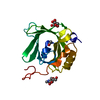
Mass: 59.044 Da / Num. of mol.: 1 / Source method: obtained synthetically / Formula: C2H3O2
#3: Sugar ChemComp-NAG / 2-acetamido-2-deoxy-beta-D-glucopyranose / N-acetyl-beta-D-glucosamine / 2-acetamido-2-deoxy-beta-D-glucose / 2-acetamido-2-deoxy-D-glucose / 2-acetamido-2-deoxy-glucose / N-ACETYL-D-GLUCOSAMINE / N-Acetylglucosamine


Type: D-saccharide, beta linking / Mass: 221.208 Da / Num. of mol.: 2
Source method: isolated from a genetically manipulated source
Formula: C8H15NO6
IdentifierTypeProgram
DGlcpNAcbCONDENSED IUPAC CARBOHYDRATE SYMBOLGMML 1.0
N-acetyl-b-D-glucopyranosamineCOMMON NAMEGMML 1.0
b-D-GlcpNAcIUPAC CARBOHYDRATE SYMBOLPDB-CARE 1.0
GlcNAcSNFG CARBOHYDRATE SYMBOLGMML 1.0
#4: Water ChemComp-HOH / water / Water


Mass: 18.015 Da / Num. of mol.: 302 / Source method: isolated from a natural source / Formula: H2O
Has ligand of interestN

-
Experimental details

-
Experiment

ExperimentMethod: X-RAY DIFFRACTION / Number of used crystals: 1

-
Sample preparation

CrystalDensity Matthews: 3.06 Å3/Da / Density % sol: 59.83 % / Description: bipyramid
Crystal growTemperature: 293 K / Method: vapor diffusion, hanging drop / pH: 5
Details: 0.2 M ammonium sulfate, 0.1 M sodium acetate (pH 5.0), 28-32% (w/v) PEG monomethyl ether 2000, 0.3 M NDSB-195

-
Data collection

DiffractionMean temperature: 100 K / Ambient temp details: N2 / Serial crystal experiment: N
Diffraction sourceSource: SYNCHROTRON / Site: Photon Factory / Beamline: BL-1A / Wavelength: 1 Å
DetectorType: DECTRIS PILATUS 2M / Detector: PIXEL / Date: May 20, 2016
RadiationMonochromator: SI(111) / Protocol: SINGLE WAVELENGTH / Monochromatic (M) / Laue (L): M / Scattering type: x-ray
Radiation wavelengthWavelength: 1 Å / Relative weight: 1
ReflectionResolution: 1.5→50.44 Å / Num. obs: 35572 / % possible obs: 100 % / Redundancy: 10.4 % / Rmerge(I) obs: 0.046 / Net I/σ(I): 26.5
Reflection shellResolution: 1.6→1.69 Å / Redundancy: 11.6 % / Rmerge(I) obs: 0.229 / Mean I/σ(I) obs: 9.5 / Num. unique obs: 4460 / CC1/2: 0.989 / Rpim(I) all: 0.073 / Rrim(I) all: 0.229 / Rsym value: 0.251 / % possible all: 100

-
Processing

Software
NameVersionClassification
REFMAC5.8.0158refinement
XDS0.48data reduction
SCALA3.3.22data scaling
SHELXCD2015/2phasing
RefinementMethod to determine structure: SAD / Resolution: 1.5→50.44 Å / Cor.coef. Fo:Fc: 0.967 / Cor.coef. Fo:Fc free: 0.958 / SU B: 1.114 / SU ML: 0.04 / Cross valid method: THROUGHOUT / ESU R: 0.063 / ESU R Free: 0.061 / Details: HYDROGENS HAVE BEEN ADDED IN THE RIDING POSITIONS
RfactorNum. reflection% reflectionSelection details
Rfree0.18686 1843 4.9 %RANDOM
Rwork0.17392 ---
obs0.17456 35572 99.97 %-
Solvent computationIon probe radii: 0.8 Å / Shrinkage radii: 0.8 Å / VDW probe radii: 1.2 Å
Displacement parametersBiso mean: 18.32 Å2
Baniso -1Baniso -2Baniso -3
1-0.44 Å20.22 Å20 Å2
2--0.44 Å20 Å2
3----1.43 Å2
Refinement stepCycle: 1 / Resolution: 1.5→50.44 Å
ProteinNucleic acidLigandSolventTotal
Num. atoms1280 0 32 305 1617
Refine LS restraints
Refine-IDTypeDev idealDev ideal targetNumber
X-RAY DIFFRACTIONr_bond_refined_d0.0070.0191390
X-RAY DIFFRACTIONr_bond_other_d0.0020.021167
X-RAY DIFFRACTIONr_angle_refined_deg1.2561.961906
X-RAY DIFFRACTIONr_angle_other_deg0.90332745
X-RAY DIFFRACTIONr_dihedral_angle_1_deg6.7165171
X-RAY DIFFRACTIONr_dihedral_angle_2_deg34.76625.65269
X-RAY DIFFRACTIONr_dihedral_angle_3_deg12.01515212
X-RAY DIFFRACTIONr_dihedral_angle_4_deg11.068153
X-RAY DIFFRACTIONr_chiral_restr0.0750.2206
X-RAY DIFFRACTIONr_gen_planes_refined0.0050.0211579
X-RAY DIFFRACTIONr_gen_planes_other0.0020.02270
X-RAY DIFFRACTIONr_nbd_refined
X-RAY DIFFRACTIONr_nbd_other
X-RAY DIFFRACTIONr_nbtor_refined
X-RAY DIFFRACTIONr_nbtor_other
X-RAY DIFFRACTIONr_xyhbond_nbd_refined
X-RAY DIFFRACTIONr_xyhbond_nbd_other
X-RAY DIFFRACTIONr_metal_ion_refined
X-RAY DIFFRACTIONr_metal_ion_other
X-RAY DIFFRACTIONr_symmetry_vdw_refined
X-RAY DIFFRACTIONr_symmetry_vdw_other
X-RAY DIFFRACTIONr_symmetry_hbond_refined
X-RAY DIFFRACTIONr_symmetry_hbond_other
X-RAY DIFFRACTIONr_symmetry_metal_ion_refined
X-RAY DIFFRACTIONr_symmetry_metal_ion_other
X-RAY DIFFRACTIONr_mcbond_it0.7051.606667
X-RAY DIFFRACTIONr_mcbond_other0.7051.605665
X-RAY DIFFRACTIONr_mcangle_it1.1462.411837
X-RAY DIFFRACTIONr_mcangle_other1.1462.411838
X-RAY DIFFRACTIONr_scbond_it1.2211.807723
X-RAY DIFFRACTIONr_scbond_other1.221.811724
X-RAY DIFFRACTIONr_scangle_it
X-RAY DIFFRACTIONr_scangle_other1.9642.6691068
X-RAY DIFFRACTIONr_long_range_B_refined4.85422.6361682
X-RAY DIFFRACTIONr_long_range_B_other4.40320.4771571
X-RAY DIFFRACTIONr_rigid_bond_restr
X-RAY DIFFRACTIONr_sphericity_free
X-RAY DIFFRACTIONr_sphericity_bonded
LS refinement shellResolution: 1.5→1.539 Å / Total num. of bins used: 20
RfactorNum. reflection% reflection
Rfree0.267 148 -
Rwork0.281 2555 -
obs--99.93 %

+
About Yorodumi

-
News

-
Feb 9, 2022. New format data for meta-information of EMDB entries

New format data for meta-information of EMDB entries

  • Version 3 of the EMDB header file is now the official format.
  • The previous official version 1.9 will be removed from the archive.

Related info.:EMDB header

External links:wwPDB to switch to version 3 of the EMDB data model

-
Aug 12, 2020. Covid-19 info

Covid-19 info

URL: https://pdbj.org/emnavi/covid19.php

New page: Covid-19 featured information page in EM Navigator.

Related info.:Covid-19 info / Mar 5, 2020. Novel coronavirus structure data

+
Mar 5, 2020. Novel coronavirus structure data

Novel coronavirus structure data

Related info.:Yorodumi Speices / Aug 12, 2020. Covid-19 info

External links:COVID-19 featured content - PDBj / Molecule of the Month (242):Coronavirus Proteases

+
Jan 31, 2019. EMDB accession codes are about to change! (news from PDBe EMDB page)

EMDB accession codes are about to change! (news from PDBe EMDB page)

  • The allocation of 4 digits for EMDB accession codes will soon come to an end. Whilst these codes will remain in use, new EMDB accession codes will include an additional digit and will expand incrementally as the available range of codes is exhausted. The current 4-digit format prefixed with “EMD-” (i.e. EMD-XXXX) will advance to a 5-digit format (i.e. EMD-XXXXX), and so on. It is currently estimated that the 4-digit codes will be depleted around Spring 2019, at which point the 5-digit format will come into force.
  • The EM Navigator/Yorodumi systems omit the EMD- prefix.

Related info.:Q: What is EMD? / ID/Accession-code notation in Yorodumi/EM Navigator

External links:EMDB Accession Codes are Changing Soon! / Contact to PDBj

+
Jul 12, 2017. Major update of PDB

Major update of PDB

  • wwPDB released updated PDB data conforming to the new PDBx/mmCIF dictionary.
  • This is a major update changing the version number from 4 to 5, and with Remediation, in which all the entries are updated.
  • In this update, many items about electron microscopy experimental information are reorganized (e.g. em_software).
  • Now, EM Navigator and Yorodumi are based on the updated data.

External links:wwPDB Remediation / Enriched Model Files Conforming to OneDep Data Standards Now Available in the PDB FTP Archive

-
Yorodumi

Thousand views of thousand structures

  • Yorodumi is a browser for structure data from EMDB, PDB, SASBDB, etc.
  • This page is also the successor to EM Navigator detail page, and also detail information page/front-end page for Omokage search.
  • The word "yorodu" (or yorozu) is an old Japanese word meaning "ten thousand". "mi" (miru) is to see.

Related info.:EMDB / PDB / SASBDB / Comparison of 3 databanks / Yorodumi Search / Aug 31, 2016. New EM Navigator & Yorodumi / Yorodumi Papers / Jmol/JSmol / Function and homology information / Changes in new EM Navigator and Yorodumi

Read more