Mass: 18.015 Da / Num. of mol.: 15 / Source method: isolated from a natural source / Formula: H2O
Has protein modification
Y
Sequence details
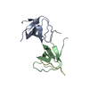
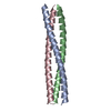
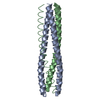
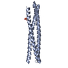
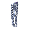
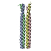
THE CRYSTALLIZED PROTEIN INCLUDES ONLY THE COILED-COIL DOMAIN OF BETA-PIX AND AS A CLONING ARTIFACT ...THE CRYSTALLIZED PROTEIN INCLUDES ONLY THE COILED-COIL DOMAIN OF BETA-PIX AND AS A CLONING ARTIFACT THE N-TERMINAL RESIDUES GPLGS.
-
Experimental details
-
Experiment
Experiment
Method: X-RAY DIFFRACTION / Number of used crystals: 1
-
Sample preparation
Crystal
Density Matthews: 3.3 Å3/Da / Density % sol: 62 % Description: RESIDUES 230 TO 278 OF CHAIN C OF PDB ID 2BA2 WERE USED FOR MR. SEQUENCE ASSIGNMENT WAS CONFIRMED USING ANOMALOUS DIFFERENCE MAPS TO VERIFY THE PRESENCE OF THE SELENIUM SITES.
Crystal grow
Details: 35% MPD, 0.24 M NON-DETERGENT SULPHOBETAINE 195 (NDSB-195)
Resolution: 2.8→39.72 Å / Cor.coef. Fo:Fc: 0.91 / Cor.coef. Fo:Fc free: 0.883 / Cross valid method: THROUGHOUT / ESU R: 0.94 / ESU R Free: 0.4 / Stereochemistry target values: MAXIMUM LIKELIHOOD Details: HYDROGENS HAVE BEEN ADDED IN THE RIDING POSITIONS. THE BIOLOGICALLY RELEVANT MOLECULE IS ARRANGED AROUND A NON-CRYSTALLOGRAPHIC THREEFOLD AXIS.
Rfactor
Num. reflection
% reflection
Selection details
Rfree
0.305
95
4.5 %
RANDOM
Rwork
0.278
-
-
-
obs
0.279
1993
99.4 %
-
Solvent computation
Ion probe radii: 0.8 Å / Shrinkage radii: 0.8 Å / VDW probe radii: 1.2 Å / Solvent model: MASK
 Movie
Movie Controller


 Open data
 Basic information
 Components
 Keywords
 Function and homology information
 X-RAY DIFFRACTION /
  Authors
 Citation
 Structure visualization
 Downloads & links
 Other downloads
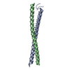


 PDBj
PDBj

 Assembly

 Mass: 18.015 Da / Num. of mol.: 15 / Source method: isolated from a natural source / Formula: H2O
 Sample preparation
 / Beamline: I04 / Wavelength: 0.9698
  Processing