 Movie
Movie Controller

[English] 日本語
 Yorodumi
- PDB-6r2g: Crystal structure of a single-chain protein mimetic of the gp41 N... -
+ Open data

- Basic information
| Entry | Database: PDB / ID: 6r2g | |||||||||
|---|---|---|---|---|---|---|---|---|---|---|
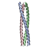
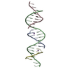
| Title | Crystal structure of a single-chain protein mimetic of the gp41 NHR trimer in complex with the synthetic CHR peptide C34 | |||||||||
 Components Components |
| |||||||||
 Keywords Keywords | BIOSYNTHETIC PROTEIN / gp41 / HIV / coiled-coil | |||||||||
| Function / homology |  Function and homology information Function and homology informationDectin-2 family / symbiont-mediated perturbation of host defense response / positive regulation of plasma membrane raft polarization / positive regulation of receptor clustering / host cell endosome membrane / clathrin-dependent endocytosis of virus by host cell / viral protein processing / fusion of virus membrane with host plasma membrane / fusion of virus membrane with host endosome membrane / viral envelope ...Dectin-2 family / symbiont-mediated perturbation of host defense response / positive regulation of plasma membrane raft polarization / positive regulation of receptor clustering / host cell endosome membrane / clathrin-dependent endocytosis of virus by host cell / viral protein processing / fusion of virus membrane with host plasma membrane / fusion of virus membrane with host endosome membrane / viral envelope / virion attachment to host cell / host cell plasma membrane / virion membrane / structural molecule activity / membrane Similarity search - Function | |||||||||
| Biological species |   Human immunodeficiency virus 1Human immunodeficiency virus type 1 group M subtype B Human immunodeficiency virus 1Human immunodeficiency virus type 1 group M subtype B | |||||||||
| Method |  X-RAY DIFFRACTION / SYNCHROTRON / MOLECULAR REPLACEMENT / molecular replacement / Resolution: 1.9 Å X-RAY DIFFRACTION / SYNCHROTRON / MOLECULAR REPLACEMENT / molecular replacement / Resolution: 1.9 Å | |||||||||
 Authors Authors | Camara-Artigas, A. / Conejero-Lara, F. / Jurado, S. / Cano-Munoz, M. / Morel, B. | |||||||||
| Funding support |  Spain, 2items Spain, 2items
| |||||||||
 Citation Citation | Journal: J.Mol.Biol. / Year: 2019 Title: Structural and Thermodynamic Analysis of HIV-1 Fusion Inhibition Using Small gp41 Mimetic Proteins. Authors: Jurado, S. / Cano-Munoz, M. / Morel, B. / Standoli, S. / Santarossa, E. / Moog, C. / Schmidt, S. / Laumond, G. / Camara-Artigas, A. / Conejero-Lara, F. #1: Journal: Proc.Natl.Acad.Sci.USA / Year: 2014Title: Single-chain protein mimetics of the N-terminal heptad-repeat region of gp41 with potential as anti-HIV-1 drugs. Authors: Crespillo, S. / Camara-Artigas, A. / Casares, S. / Morel, B. / Cobos, E.S. / Mateo, P.L. / Mouz, N. / Martin, C.E. / Roger, M.G. / El Habib, R. / Su, B. / Moog, C. / Conejero-Lara, F. | |||||||||
| History |
|
- Structure visualization
| Structure viewer | Molecule: MolmilJmol/JSmol |
|---|
- Downloads & links
-Download
| PDBx/mmCIF format | 6r2g.cif.gz | 120.2 KB | Display | PDBx/mmCIF format |
|---|---|---|---|---|
| PDB format | pdb6r2g.ent.gz | 94.9 KB | Display | PDB format |
| PDBx/mmJSON format | 6r2g.json.gz | Tree view | PDBx/mmJSON format | |
| Others |  Other downloads Other downloads |
-Validation report
| Arichive directory | https://data.pdbj.org/pub/pdb/validation_reports/r2/6r2gftp://data.pdbj.org/pub/pdb/validation_reports/r2/6r2g | HTTPS FTP |
|---|
-Related structure data
| Related structure data | 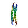 4r61S S: Starting model for refinement |
|---|---|
| Similar structure data |


-Links
 PDBj
PDBj


- Assembly
| Deposited unit | 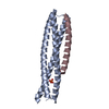
| ||||||||
|---|---|---|---|---|---|---|---|---|---|
| 1 | 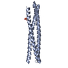
| ||||||||
| 2 | 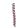
| ||||||||
| Unit cell |
|
-Components
| #1: Protein | Mass: 21176.070 Da / Num. of mol.: 1 Source method: isolated from a genetically manipulated source Details: Single-chain protein mimetics of the N-terminal heptad-repeat region of gp41 Source: (gene. exp.) Human immunodeficiency virus 1 / Production host:  |
|---|---|
| #2: Protein/peptide | Mass: 4275.623 Da / Num. of mol.: 1 Source method: isolated from a genetically manipulated source Source: (gene. exp.) Human immunodeficiency virus type 1 group M subtype B (isolate BRU/LAI)Strain: isolate BRU/LAI / Gene: env / Production host: |
| #3: Chemical | ChemComp-PO4 /   Mass: 94.971 Da / Num. of mol.: 1 / Source method: obtained synthetically / Formula: PO4 Mass: 94.971 Da / Num. of mol.: 1 / Source method: obtained synthetically / Formula: PO4 |
| #4: Water | ChemComp-HOH /  Mass: 18.015 Da / Num. of mol.: 53 / Source method: isolated from a natural source / Formula: H2O Mass: 18.015 Da / Num. of mol.: 53 / Source method: isolated from a natural source / Formula: H2O |
| Has protein modification | Y |
-Experimental details
-Experiment
| Experiment | Method: X-RAY DIFFRACTION / Number of used crystals: 1 |
|---|
- Sample preparation
| Crystal | Density Matthews: 1.78 Å3/Da / Density % sol: 31.04 % |
|---|---|
| Crystal grow | Temperature: 298 K / Method: vapor diffusion, sitting drop / pH: 6.5 / Details: 20% PEG 4000, 100 mM MES |
-Data collection
| Diffraction | Mean temperature: 100 K / Serial crystal experiment: N | ||||||||||||||||||||||||
|---|---|---|---|---|---|---|---|---|---|---|---|---|---|---|---|---|---|---|---|---|---|---|---|---|---|
| Diffraction source | Source: SYNCHROTRON / Site: ESRF  / Beamline: ID30B / Wavelength: 0.97625 Å / Beamline: ID30B / Wavelength: 0.97625 Å | ||||||||||||||||||||||||
| Detector | Type: DECTRIS PILATUS3 6M / Detector: PIXEL / Date: Jan 11, 2018 | ||||||||||||||||||||||||
| Radiation | Protocol: SINGLE WAVELENGTH / Monochromatic (M) / Laue (L): M / Scattering type: x-ray | ||||||||||||||||||||||||
| Radiation wavelength | Wavelength: 0.97625 Å / Relative weight: 1 | ||||||||||||||||||||||||
| Reflection | Resolution: 1.9→19.85 Å / Num. obs: 15041 / % possible obs: 99.8 % / Redundancy: 5.1 % / CC1/2: 0.999 / Rmerge(I) obs: 0.059 / Rpim(I) all: 0.029 / Rrim(I) all: 0.066 / Net I/σ(I): 12.9 | ||||||||||||||||||||||||
| Reflection shell | Diffraction-ID: 1
|
-Phasing
| Phasing | Method: molecular replacement |
|---|
- Processing
| Software |
| |||||||||||||||||||||||||||||||||||||||||||||||||||||||||||||||||||||||||||||||||||||||||||||||||||||||||||||||||||||||||||||
|---|---|---|---|---|---|---|---|---|---|---|---|---|---|---|---|---|---|---|---|---|---|---|---|---|---|---|---|---|---|---|---|---|---|---|---|---|---|---|---|---|---|---|---|---|---|---|---|---|---|---|---|---|---|---|---|---|---|---|---|---|---|---|---|---|---|---|---|---|---|---|---|---|---|---|---|---|---|---|---|---|---|---|---|---|---|---|---|---|---|---|---|---|---|---|---|---|---|---|---|---|---|---|---|---|---|---|---|---|---|---|---|---|---|---|---|---|---|---|---|---|---|---|---|---|---|---|
| Refinement | Method to determine structure: MOLECULAR REPLACEMENT Starting model: 4R61 Resolution: 1.9→19.852 Å / SU ML: 0.21 / Cross valid method: THROUGHOUT / σ(F): 0.21 / Phase error: 26.76 / Stereochemistry target values: ML
| |||||||||||||||||||||||||||||||||||||||||||||||||||||||||||||||||||||||||||||||||||||||||||||||||||||||||||||||||||||||||||||
| Solvent computation | Shrinkage radii: 0.9 Å / VDW probe radii: 1.11 Å / Solvent model: FLAT BULK SOLVENT MODEL | |||||||||||||||||||||||||||||||||||||||||||||||||||||||||||||||||||||||||||||||||||||||||||||||||||||||||||||||||||||||||||||
| Refinement step | Cycle: LAST / Resolution: 1.9→19.852 Å
| |||||||||||||||||||||||||||||||||||||||||||||||||||||||||||||||||||||||||||||||||||||||||||||||||||||||||||||||||||||||||||||
| Refine LS restraints |
| |||||||||||||||||||||||||||||||||||||||||||||||||||||||||||||||||||||||||||||||||||||||||||||||||||||||||||||||||||||||||||||
| LS refinement shell |
| |||||||||||||||||||||||||||||||||||||||||||||||||||||||||||||||||||||||||||||||||||||||||||||||||||||||||||||||||||||||||||||
| Refinement TLS params. | Method: refined / Refine-ID: X-RAY DIFFRACTION
| |||||||||||||||||||||||||||||||||||||||||||||||||||||||||||||||||||||||||||||||||||||||||||||||||||||||||||||||||||||||||||||
| Refinement TLS group |
|
