 Movie
Movie Controller

[English] 日本語
 Yorodumi
- PDB-4r61: Crystal structure of a rationally designed single-chain protein m... -
+ Open data

- Basic information
| Entry | Database: PDB / ID: 4r61 | ||||||
|---|---|---|---|---|---|---|---|
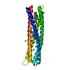
| Title | Crystal structure of a rationally designed single-chain protein mimicking a trimeric gp41 N-terminal heptad-repeat region | ||||||
 Components Components | gp41-based construct covNHR3-ABC | ||||||
 Keywords Keywords | VIRAL PROTEIN / coiled-coil | ||||||
| Biological species |   Human immunodeficiency virus 1 Human immunodeficiency virus 1 | ||||||
| Method |  X-RAY DIFFRACTION / SYNCHROTRON / MOLECULAR REPLACEMENT / molecular replacement / Resolution: 3.1 Å X-RAY DIFFRACTION / SYNCHROTRON / MOLECULAR REPLACEMENT / molecular replacement / Resolution: 3.1 Å | ||||||
 Authors Authors | Camara-Artigas, A. / Conejero-Lara, F. / Crespillo, S. / Casares, S. | ||||||
 Citation Citation | Journal: Proc.Natl.Acad.Sci.USA / Year: 2014 Title: Single-chain protein mimetics of the N-terminal heptad-repeat region of gp41 with potential as anti-HIV-1 drugs. Authors: Crespillo, S. / Camara-Artigas, A. / Casares, S. / Morel, B. / Cobos, E.S. / Mateo, P.L. / Mouz, N. / Martin, C.E. / Roger, M.G. / El Habib, R. / Su, B. / Moog, C. / Conejero-Lara, F. | ||||||
| History |
|
- Structure visualization
| Structure viewer | Molecule:  MolmilJmol/JSmol MolmilJmol/JSmol |
|---|
- Downloads & links
-Download
| PDBx/mmCIF format | 4r61.cif.gz | 40.5 KB | Display | PDBx/mmCIF format |
|---|---|---|---|---|
| PDB format | pdb4r61.ent.gz | 28 KB | Display | PDB format |
| PDBx/mmJSON format | 4r61.json.gz | Tree view | PDBx/mmJSON format | |
| Others |  Other downloads Other downloads |
-Validation report
| Arichive directory | https://data.pdbj.org/pub/pdb/validation_reports/r6/4r61ftp://data.pdbj.org/pub/pdb/validation_reports/r6/4r61 | HTTPS FTP |
|---|
-Related structure data
| Related structure data | 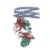 2xraS S: Starting model for refinement |
|---|---|
| Similar structure data |


-Links
 PDBj
PDBj- Assembly
| Deposited unit | 
| ||||||||
|---|---|---|---|---|---|---|---|---|---|
| 1 |
| ||||||||
| Unit cell |
|
-Components
| #1: Protein | Mass: 20265.139 Da / Num. of mol.: 1 Source method: isolated from a genetically manipulated source Source: (gene. exp.) Human immunodeficiency virus 1 / Production host:  |
|---|
-Experimental details
-Experiment
| Experiment | Method: X-RAY DIFFRACTION / Number of used crystals: 1 |
|---|
- Sample preparation
| Crystal | Density Matthews: 2.05 Å3/Da / Density % sol: 40.14 % |
|---|---|
| Crystal grow | Temperature: 298 K / Method: vapor diffusion, sitting drop / pH: 4.6 Details: 2 M sodium chloride, 0.1 M sodium acetate trihydrate, pH 4.6, VAPOR DIFFUSION, SITTING DROP, temperature 298K |
-Data collection
| Diffraction |
| |||||||||||||||||||||
|---|---|---|---|---|---|---|---|---|---|---|---|---|---|---|---|---|---|---|---|---|---|---|
| Diffraction source |
| |||||||||||||||||||||
| Detector |
| |||||||||||||||||||||
| Radiation |
| |||||||||||||||||||||
| Radiation wavelength |
| |||||||||||||||||||||
| Reflection | Redundancy: 8.6 % / Number: 28217 / Rmerge(I) obs: 0.09 / D res high: 3.1 Å / D res low: 19.8 Å / Num. obs: 3278 / % possible obs: 99.6 / Rejects: 634 | |||||||||||||||||||||
| Diffraction reflection shell |
| |||||||||||||||||||||
| Reflection | Resolution: 3.1→19.8 Å / Num. all: 5202 / Num. obs: 3278 / % possible obs: 99.6 % / Observed criterion σ(F): 0 / Observed criterion σ(I): 0 / Redundancy: 8.6 % / Biso Wilson estimate: 44.52 Å2 / Rmerge(I) obs: 0.09 / Net I/σ(I): 130.8 / Scaling rejects: 634 | |||||||||||||||||||||
| Reflection shell | Diffraction-ID: 1,2
|


-Phasing
| Phasing | Method: molecular replacement |
|---|
- Processing
| Software |
| ||||||||||||||||||||||||||||
|---|---|---|---|---|---|---|---|---|---|---|---|---|---|---|---|---|---|---|---|---|---|---|---|---|---|---|---|---|---|
| Refinement | Method to determine structure: MOLECULAR REPLACEMENT Starting model: PDB ENTRY 2XRA Resolution: 3.1→17.803 Å / SU ML: 0.31 / Cross valid method: THROUGHOUT / σ(F): 1.91 / σ(I): 0 / Phase error: 30.83 / Stereochemistry target values: ML
| ||||||||||||||||||||||||||||
| Solvent computation | Shrinkage radii: 0.9 Å / VDW probe radii: 1.11 Å / Solvent model: FLAT BULK SOLVENT MODEL | ||||||||||||||||||||||||||||
| Displacement parameters | Biso max: 198.97 Å2 / Biso mean: 72.45 Å2 / Biso min: 12.99 Å2 | ||||||||||||||||||||||||||||
| Refinement step | Cycle: LAST / Resolution: 3.1→17.803 Å
| ||||||||||||||||||||||||||||
| Refine LS restraints |
| ||||||||||||||||||||||||||||
| LS refinement shell | Refine-ID: X-RAY DIFFRACTION / Total num. of bins used: 2
|
