[English] 日本語
Yorodumi
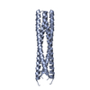
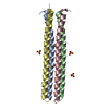
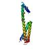
- PDB-5h1x: Crystal Structure of rat Nup62 Coiled-coil motif -

+
Open data


ID or keywords:

Loading...

-
Basic information

Entry
Database: PDB / ID: 5h1x
TitleCrystal Structure of rat Nup62 Coiled-coil motif
ComponentsNuclear pore glycoprotein p62
KeywordsSTRUCTURAL PROTEIN / coiled-coil
Function / homology
Function and homology information


Transport of the SLBP independent Mature mRNA / Transport of the SLBP Dependant Mature mRNA / Transport of Mature mRNA Derived from an Intronless Transcript / Transport of Mature mRNA derived from an Intron-Containing Transcript / snRNP Assembly / SUMOylation of ubiquitinylation proteins / Nuclear Pore Complex (NPC) Disassembly / SUMOylation of SUMOylation proteins / SUMOylation of chromatin organization proteins / SUMOylation of RNA binding proteins ...Transport of the SLBP independent Mature mRNA / Transport of the SLBP Dependant Mature mRNA / Transport of Mature mRNA Derived from an Intronless Transcript / Transport of Mature mRNA derived from an Intron-Containing Transcript / snRNP Assembly / SUMOylation of ubiquitinylation proteins / Nuclear Pore Complex (NPC) Disassembly / SUMOylation of SUMOylation proteins / SUMOylation of chromatin organization proteins / SUMOylation of RNA binding proteins / SUMOylation of DNA replication proteins / Transcriptional regulation by small RNAs / Regulation of Glucokinase by Glucokinase Regulatory Protein / SUMOylation of DNA damage response and repair proteins / centriole assembly / regulation of protein import into nucleus / positive regulation of centriole replication / positive regulation of mitotic cytokinetic process / annulate lamellae / Regulation of HSF1-mediated heat shock response / nuclear pore central transport channel / positive regulation of protein localization to centrosome / negative regulation of Ras protein signal transduction / structural constituent of nuclear pore / Flemming body / negative regulation of programmed cell death / mitotic centrosome separation / RNA export from nucleus / centrosome cycle / nuclear thyroid hormone receptor binding / PTB domain binding / mitotic metaphase chromosome alignment / negative regulation of epidermal growth factor receptor signaling pathway / kinesin binding / mRNA transport / nuclear pore / regulation of mitotic spindle organization / Hsp70 protein binding / SH2 domain binding / positive regulation of mitotic nuclear division / ubiquitin binding / Hsp90 protein binding / mitotic spindle / phospholipid binding / spindle pole / protein import into nucleus / cellular senescence / signaling receptor complex adaptor activity / nuclear envelope / spermatogenesis / nuclear membrane / positive regulation of canonical NF-kappaB signal transduction / cell surface receptor signaling pathway / ribonucleoprotein complex / negative regulation of cell population proliferation / centrosome / protein-containing complex binding / negative regulation of apoptotic process / positive regulation of DNA-templated transcription / protein-containing complex / cytoplasm
Similarity search - Function
Nucleoporin, NSP1-like, C-terminal / Nucleoporin NSP1/NUP62 / Nsp1-like C-terminal region
Similarity search - Domain/homology
Nuclear pore glycoprotein p62
Similarity search - Component
Biological speciesRattus norvegicus (Norway rat)
MethodX-RAY DIFFRACTION / SYNCHROTRON / MOLECULAR REPLACEMENT / Resolution: 2.41 Å
Model details362-425 fragment
AuthorsPravin, D.
Funding support India, 1items
OrganizationGrant numberCountry
DST, DBT India
CitationJournal: Biochemistry / Year: 2017
Title: The Nup62 Coiled-Coil Motif Provides Plasticity for Triple-Helix Bundle Formation
Authors: Dewangan, P.S. / Sonawane, P.J. / Chouksey, A.R. / Chauhan, R.
History
DepositionOct 12, 2016Deposition site: PDBJ / Processing site: PDBJ
Revision 1.0Oct 4, 2017Provider: repository / Type: Initial release
Revision 1.1Nov 8, 2023Group: Data collection / Database references / Refinement description
Category: chem_comp_atom / chem_comp_bond ...chem_comp_atom / chem_comp_bond / database_2 / pdbx_initial_refinement_model
Item: _database_2.pdbx_DOI / _database_2.pdbx_database_accession

-
Structure visualization

Structure viewerMolecule:
MolmilJmol/JSmol

Downloads & links

-
Assembly

Deposited unit
A: Nuclear pore glycoprotein p62
B: Nuclear pore glycoprotein p62
C: Nuclear pore glycoprotein p62


Theoretical massNumber of molelcules
Total (without water)18,6093
Polymers18,6093
Non-polymers00
Water19811
1


  • Idetical with deposited unit
  • defined by author&software
TypeNameSymmetry operationNumber
identity operation1_555x,y,z1
Buried area4890 Å2
ΔGint-38 kcal/mol
Surface area10370 Å2
MethodPISA
Unit cell
Length a, b, c (Å)30.982, 97.563, 31.502
Angle α, β, γ (deg.)90.000, 112.850, 90.000
Int Tables number4
Space group name H-MP1211

-
Components

#1: Protein Nuclear pore glycoprotein p62 / 62 kDa nucleoporin / Nucleoporin Nup62


Mass: 6203.017 Da / Num. of mol.: 3 / Fragment: UNP residues 362-412
Source method: isolated from a genetically manipulated source
Source: (gene. exp.) Rattus norvegicus (Norway rat) / Gene: Nup62 / Plasmid: pET28a / Production host: Escherichia coli (E. coli) / Strain (production host): BL21(RIL) / References: UniProt: P17955
#2: Water ChemComp-HOH / water


Mass: 18.015 Da / Num. of mol.: 11 / Source method: isolated from a natural source / Formula: H2O

-
Experimental details

-
Experiment

ExperimentMethod: X-RAY DIFFRACTION / Number of used crystals: 1

-
Sample preparation

CrystalDensity Matthews: 2.36 Å3/Da / Density % sol: 47.83 %
Description: THE ENTRY CONTAINS FRIEDEL PAIRS IN F_PLUS/MINUS COLUMNS.
Crystal growTemperature: 291 K / Method: evaporation / pH: 8
Details: 100mM Tris-HCl, 50mM Magnesium Chloride, 34% PEG 400
PH range: 8.0-8.5

-
Data collection

DiffractionMean temperature: 100 K
Diffraction sourceSource: SYNCHROTRON / Site: ELETTRA / Beamline: 5.2R / Wavelength: 0.98 Å
DetectorType: DECTRIS PILATUS 2M / Detector: PIXEL / Date: Dec 16, 2013
RadiationProtocol: SINGLE WAVELENGTH / Monochromatic (M) / Laue (L): M / Scattering type: x-ray
Radiation wavelengthWavelength: 0.98 Å / Relative weight: 1
ReflectionResolution: 2.408→48.782 Å / Num. all: 6669 / Num. obs: 6669 / % possible obs: 99.4 % / Redundancy: 3.3 % / Biso Wilson estimate: 41.49 Å2 / Rpim(I) all: 0.025 / Rrim(I) all: 0.047 / Rsym value: 0.039 / Net I/av σ(I): 8.448 / Net I/σ(I): 17.9 / Num. measured all: 22313
Reflection shell

Diffraction-ID: 1 / Rejects: _

Resolution (Å)Redundancy (%)Rmerge(I) obsMean I/σ(I) obsNum. measured allNum. unique allRpim(I) allRrim(I) allRsym valueNet I/σ(I) obs% possible all
2.41-2.543.20.1764.330349340.1140.2110.1765.998.6
2.54-2.693.50.1365.732579330.0860.1620.1367.899.9
2.69-2.883.50.17.630518830.0640.1190.110.299.9
2.88-3.113.40.07110.227078010.0460.0840.07113.399.6
3.11-3.413.20.0514.423677430.0330.0610.0518.599.7
3.41-3.813.30.03817.521906680.0250.0450.03825.799.6
3.81-4.43.50.0318.720775990.0190.0350.0333.299.5
4.4-5.393.30.0291916474960.0190.0350.02933.499.3
5.39-7.623.20.03218.112393900.0210.0380.03228.298.6
7.62-48.7823.40.02613.17442220.0170.0320.02638.598.9

-
Processing

Software
NameVersionClassification
PHENIX1.8.4_1496refinement
SCALAdata scaling
PDB_EXTRACT3.2data extraction
XDSdata reduction
PHENIX1.8.4_1496phasing
RefinementMethod to determine structure: MOLECULAR REPLACEMENT
Starting model: 3T97
Resolution: 2.41→27.825 Å / FOM work R set: 0.7039 / SU ML: 0.3 / Cross valid method: FREE R-VALUE / σ(F): 1.34 / Phase error: 35.51 / Stereochemistry target values: ML
RfactorNum. reflection% reflection
Rfree0.3077 662 9.99 %
Rwork0.2269 5965 -
obs0.2351 6627 99.33 %
Solvent computationShrinkage radii: 0.9 Å / VDW probe radii: 1.11 Å / Solvent model: FLAT BULK SOLVENT MODEL
Displacement parametersBiso max: 98.21 Å2 / Biso mean: 54.65 Å2 / Biso min: 29.02 Å2
Refinement stepCycle: final / Resolution: 2.41→27.825 Å
ProteinNucleic acidLigandSolventTotal
Num. atoms1228 0 0 11 1239
Biso mean---53.41 -
Num. residues----146
Refine LS restraints
Refine-IDTypeDev idealNumber
X-RAY DIFFRACTIONf_bond_d0.0081238
X-RAY DIFFRACTIONf_angle_d1.121657
X-RAY DIFFRACTIONf_chiral_restr0.052187
X-RAY DIFFRACTIONf_plane_restr0.006216
X-RAY DIFFRACTIONf_dihedral_angle_d18.675496
LS refinement shell

Refine-ID: X-RAY DIFFRACTION / Total num. of bins used: 5

Resolution (Å)Rfactor RfreeNum. reflection RfreeRfactor RworkNum. reflection RworkNum. reflection all% reflection obs (%)
2.4102-2.59620.31541330.22921180131399
2.5962-2.85720.3361350.233812031338100
2.8572-3.27010.37761300.2621165129599
3.2701-4.11780.29141270.224612121339100
4.1178-27.82670.28361370.21331205134299

+
About Yorodumi

-
News

-
Feb 9, 2022. New format data for meta-information of EMDB entries

New format data for meta-information of EMDB entries

  • Version 3 of the EMDB header file is now the official format.
  • The previous official version 1.9 will be removed from the archive.

Related info.:EMDB header

External links:wwPDB to switch to version 3 of the EMDB data model

-
Aug 12, 2020. Covid-19 info

Covid-19 info

URL: https://pdbjlvh1.pdbj.org/emnavi/covid19.php

New page: Covid-19 featured information page in EM Navigator.

Related info.:Covid-19 info / Mar 5, 2020. Novel coronavirus structure data

+
Mar 5, 2020. Novel coronavirus structure data

Novel coronavirus structure data

Related info.:Yorodumi Speices / Aug 12, 2020. Covid-19 info

External links:COVID-19 featured content - PDBj / Molecule of the Month (242):Coronavirus Proteases

+
Jan 31, 2019. EMDB accession codes are about to change! (news from PDBe EMDB page)

EMDB accession codes are about to change! (news from PDBe EMDB page)

  • The allocation of 4 digits for EMDB accession codes will soon come to an end. Whilst these codes will remain in use, new EMDB accession codes will include an additional digit and will expand incrementally as the available range of codes is exhausted. The current 4-digit format prefixed with “EMD-” (i.e. EMD-XXXX) will advance to a 5-digit format (i.e. EMD-XXXXX), and so on. It is currently estimated that the 4-digit codes will be depleted around Spring 2019, at which point the 5-digit format will come into force.
  • The EM Navigator/Yorodumi systems omit the EMD- prefix.

Related info.:Q: What is EMD? / ID/Accession-code notation in Yorodumi/EM Navigator

External links:EMDB Accession Codes are Changing Soon! / Contact to PDBj

+
Jul 12, 2017. Major update of PDB

Major update of PDB

  • wwPDB released updated PDB data conforming to the new PDBx/mmCIF dictionary.
  • This is a major update changing the version number from 4 to 5, and with Remediation, in which all the entries are updated.
  • In this update, many items about electron microscopy experimental information are reorganized (e.g. em_software).
  • Now, EM Navigator and Yorodumi are based on the updated data.

External links:wwPDB Remediation / Enriched Model Files Conforming to OneDep Data Standards Now Available in the PDB FTP Archive

-
Yorodumi

Thousand views of thousand structures

  • Yorodumi is a browser for structure data from EMDB, PDB, SASBDB, etc.
  • This page is also the successor to EM Navigator detail page, and also detail information page/front-end page for Omokage search.
  • The word "yorodu" (or yorozu) is an old Japanese word meaning "ten thousand". "mi" (miru) is to see.

Related info.:EMDB / PDB / SASBDB / Comparison of 3 databanks / Yorodumi Search / Aug 31, 2016. New EM Navigator & Yorodumi / Yorodumi Papers / Jmol/JSmol / Function and homology information / Changes in new EM Navigator and Yorodumi

Read more