 Movie
Movie Controller


+ Open data

- Basic information
| Entry | Database: PDB / ID: 6b87 | ||||||
|---|---|---|---|---|---|---|---|
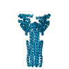
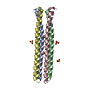
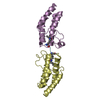
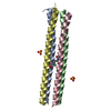
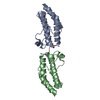
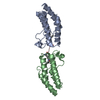
| Title | Crystal structure of transmembrane protein TMHC2_E | ||||||
 Components Components | TMHC2_E | ||||||
 Keywords Keywords | MEMBRANE PROTEIN / De novo design / transmembrane protein / helical bundle / dimer | ||||||
| Biological species | synthetic construct (others) | ||||||
| Method |  X-RAY DIFFRACTION / SYNCHROTRON / MOLECULAR REPLACEMENT / Resolution: 2.947 Å X-RAY DIFFRACTION / SYNCHROTRON / MOLECULAR REPLACEMENT / Resolution: 2.947 Å | ||||||
 Authors Authors | Lu, P. / DiMaio, F. / Min, D. / Wei, K.Y. / Bowie, J. / Baker, D. | ||||||
 Citation Citation | Journal: Science / Year: 2018 Title: Accurate computational design of multipass transmembrane proteins. Authors: Lu, P. / Min, D. / DiMaio, F. / Wei, K.Y. / Vahey, M.D. / Boyken, S.E. / Chen, Z. / Fallas, J.A. / Ueda, G. / Sheffler, W. / Mulligan, V.K. / Xu, W. / Bowie, J.U. / Baker, D. | ||||||
| History |
|
- Structure visualization
| Structure viewer | Molecule:  MolmilJmol/JSmol MolmilJmol/JSmol |
|---|
- Downloads & links
-Download
| PDBx/mmCIF format | 6b87.cif.gz | 158.6 KB | Display | PDBx/mmCIF format |
|---|---|---|---|---|
| PDB format | pdb6b87.ent.gz | 129.6 KB | Display | PDB format |
| PDBx/mmJSON format | 6b87.json.gz | Tree view | PDBx/mmJSON format | |
| Others |  Other downloads Other downloads |
-Validation report
| Arichive directory | https://data.pdbj.org/pub/pdb/validation_reports/b8/6b87ftp://data.pdbj.org/pub/pdb/validation_reports/b8/6b87 | HTTPS FTP |
|---|
-Related structure data


-Links
 PDBj
PDBj
- Assembly
| Deposited unit | 
| ||||||||
|---|---|---|---|---|---|---|---|---|---|
| 1 | 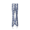
| ||||||||
| 2 | 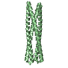
| ||||||||
| 3 | 
| ||||||||
| Unit cell |
|
-Components
| #1: Protein | Mass: 13293.877 Da / Num. of mol.: 4 Source method: isolated from a genetically manipulated source Source: (gene. exp.) synthetic construct (others) / Production host:  |
|---|
-Experimental details
-Experiment
| Experiment | Method: X-RAY DIFFRACTION / Number of used crystals: 1 |
|---|
- Sample preparation
| Crystal | Density Matthews: 2.67 Å3/Da / Density % sol: 53.86 % |
|---|---|
| Crystal grow | Temperature: 291 K / Method: vapor diffusion, hanging drop Details: 0.05 M magnesium acetate tetrahydrate, 0.05 M sodium acetate, pH 5.4, 30% v/v PEG400 |
-Data collection
| Diffraction | Mean temperature: 100 K |
|---|---|
| Diffraction source | Source: SYNCHROTRON / Site: ALS  / Beamline: 8.2.1 / Wavelength: 0.9793 Å / Beamline: 8.2.1 / Wavelength: 0.9793 Å |
| Detector | Type: ADSC QUANTUM 315r / Detector: CCD / Date: Oct 21, 2016 |
| Radiation | Monochromator: double crystal Si(111) / Protocol: SINGLE WAVELENGTH / Monochromatic (M) / Laue (L): M / Scattering type: x-ray |
| Radiation wavelength | Wavelength: 0.9793 Å / Relative weight: 1 |
| Reflection | Resolution: 2.95→50 Å / Num. obs: 10899 / % possible obs: 92.5 % / Redundancy: 3.5 % / Net I/σ(I): 9.6 |
| Reflection shell | Highest resolution: 2.95 Å |
- Processing
| Software |
| |||||||||||||||||||||||||||||||||||||||||||||||||||||||||||||||
|---|---|---|---|---|---|---|---|---|---|---|---|---|---|---|---|---|---|---|---|---|---|---|---|---|---|---|---|---|---|---|---|---|---|---|---|---|---|---|---|---|---|---|---|---|---|---|---|---|---|---|---|---|---|---|---|---|---|---|---|---|---|---|---|---|
| Refinement | Method to determine structure: MOLECULAR REPLACEMENT / Resolution: 2.947→36.942 Å / SU ML: 0.45 / Cross valid method: FREE R-VALUE / σ(F): 1.37 / Phase error: 36.18 / Stereochemistry target values: ML
| |||||||||||||||||||||||||||||||||||||||||||||||||||||||||||||||
| Solvent computation | Shrinkage radii: 0.9 Å / VDW probe radii: 1.11 Å / Solvent model: FLAT BULK SOLVENT MODEL | |||||||||||||||||||||||||||||||||||||||||||||||||||||||||||||||
| Refinement step | Cycle: LAST / Resolution: 2.947→36.942 Å
| |||||||||||||||||||||||||||||||||||||||||||||||||||||||||||||||
| Refine LS restraints |
| |||||||||||||||||||||||||||||||||||||||||||||||||||||||||||||||
| LS refinement shell |
|
