 Movie
Movie Controller

[English] 日本語
 Yorodumi
- PDB-2w40: Crystal structure of Plasmodium falciparum glycerol kinase with b... -
+ Open data

- Basic information
| Entry | Database: PDB / ID: 2w40 | ||||||
|---|---|---|---|---|---|---|---|
| Title | Crystal structure of Plasmodium falciparum glycerol kinase with bound glycerol | ||||||
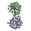
 Components Components | GLYCEROL KINASE, PUTATIVE | ||||||
 Keywords Keywords | TRANSFERASE / CLOSED CONFORMATION / KINASE / MALARIA / PLASMODIUM / SUGAR KINASE/HSP70/ACTIN SUPERFAMILY / GLYCEROL KINASE / OPEN CONFORMATION | ||||||
| Function / homology |  Function and homology information Function and homology informationTriglyceride biosynthesis / glycerol-3-phosphate biosynthetic process / glycerol kinase / glycerol kinase activity / glycerol metabolic process / glycerol catabolic process / triglyceride metabolic process / mitochondrion / ATP binding Similarity search - Function | ||||||
| Biological species |  | ||||||
| Method |  X-RAY DIFFRACTION / SYNCHROTRON / MOLECULAR REPLACEMENT / Resolution: 1.49 Å X-RAY DIFFRACTION / SYNCHROTRON / MOLECULAR REPLACEMENT / Resolution: 1.49 Å | ||||||
 Authors Authors | Schnick, C. / Polley, S.D. / Fivelman, Q.L. / Ranford-Cartwright, L. / Wilkinson, S.R. / Brannigan, J.A. / Wilkinson, A.J. / Baker, D.A. | ||||||
 Citation Citation | Journal: Mol.Microbiol. / Year: 2009 Title: Structure and Non-Essential Function of Glycerol Kinase in Plasmodium Falciparum Blood Stages. Authors: Schnick, C. / Polley, S.D. / Fivelman, Q.L. / Ranford-Cartwright, L. / Wilkinson, S.R. / Brannigan, J.A. / Wilkinson, A.J. / Baker, D.A. | ||||||
| History |
|
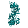
- Structure visualization
| Structure viewer | Molecule: MolmilJmol/JSmol |
|---|
- Downloads & links
-Download
| PDBx/mmCIF format | 2w40.cif.gz | 824.6 KB | Display | PDBx/mmCIF format |
|---|---|---|---|---|
| PDB format | pdb2w40.ent.gz | 683.7 KB | Display | PDB format |
| PDBx/mmJSON format | 2w40.json.gz | Tree view | PDBx/mmJSON format | |
| Others |  Other downloads Other downloads |
-Validation report
| Arichive directory | https://data.pdbj.org/pub/pdb/validation_reports/w4/2w40ftp://data.pdbj.org/pub/pdb/validation_reports/w4/2w40 | HTTPS FTP |
|---|
-Related structure data
| Related structure data | 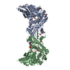 2w41C 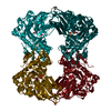 1bu6S C: citing same article ( S: Starting model for refinement |
|---|---|
| Similar structure data |
-Links
 PDBj
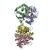
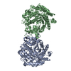
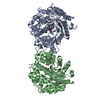
PDBj- Assembly
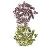
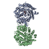
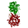
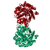
| Deposited unit | 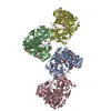
| ||||||||
|---|---|---|---|---|---|---|---|---|---|
| 1 | 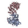
| ||||||||
| 2 | 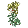
| ||||||||
| Unit cell |
|
-Components
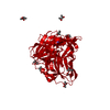
| #1: Protein | Mass: 56858.887 Da / Num. of mol.: 4 Source method: isolated from a genetically manipulated source Source: (gene. exp.) Strain: 3D7 / Plasmid: PMAL-C2X / Production host:  #2: Chemical | ChemComp-EDO /   Mass: 62.068 Da / Num. of mol.: 62 / Source method: obtained synthetically / Formula: C2H6O2 Mass: 62.068 Da / Num. of mol.: 62 / Source method: obtained synthetically / Formula: C2H6O2#3: Chemical | ChemComp-GOL /   Mass: 92.094 Da / Num. of mol.: 12 / Source method: obtained synthetically / Formula: C3H8O3 Mass: 92.094 Da / Num. of mol.: 12 / Source method: obtained synthetically / Formula: C3H8O3#4: Water | ChemComp-HOH / |  Mass: 18.015 Da / Num. of mol.: 1929 / Source method: isolated from a natural source / Formula: H2O Mass: 18.015 Da / Num. of mol.: 1929 / Source method: isolated from a natural source / Formula: H2O |
|---|
-Experimental details
-Experiment
| Experiment | Method: X-RAY DIFFRACTION / Number of used crystals: 1 |
|---|
- Sample preparation
| Crystal | Density Matthews: 2.44 Å3/Da / Density % sol: 49.6 % / Description: NONE |
|---|---|
| Crystal grow | pH: 7.5 Details: 20% PEG 3350, 50 MM KFORMATE, 25 % ETHYLENE GLYCOL, 10 MM LDAO, 20 MM GLYCEROL, pH 7.5 |
-Data collection
| Diffraction | Mean temperature: 100 K |
|---|---|
| Diffraction source | Source: SYNCHROTRON / Site: ESRF  / Beamline: ID14-2 / Wavelength: 0.933 / Beamline: ID14-2 / Wavelength: 0.933 |
| Detector | Type: ADSC CCD / Detector: CCD / Date: Jul 24, 2006 |
| Radiation | Protocol: SINGLE WAVELENGTH / Monochromatic (M) / Laue (L): M / Scattering type: x-ray |
| Radiation wavelength | Wavelength: 0.933 Å / Relative weight: 1 |
| Reflection | Resolution: 1.49→107.83 Å / Num. obs: 260542 / % possible obs: 73.5 % / Observed criterion σ(I): 2 / Redundancy: 4.3 % / Rmerge(I) obs: 0.08 / Net I/σ(I): 12 |
| Reflection shell | Resolution: 1.49→1.53 Å / Redundancy: 3.4 % / Rmerge(I) obs: 0.63 / Mean I/σ(I) obs: 2.23 / % possible all: 18 |
- Processing
| Software |
| ||||||||||||||||||||||||||||||||||||||||||||||||||||||||||||||||||||||||||||||||||||||||||||||||||||||||||||||||||||||||||||||||||||||||||||||||||||||||||||||||||||||||||||||||||||||
|---|---|---|---|---|---|---|---|---|---|---|---|---|---|---|---|---|---|---|---|---|---|---|---|---|---|---|---|---|---|---|---|---|---|---|---|---|---|---|---|---|---|---|---|---|---|---|---|---|---|---|---|---|---|---|---|---|---|---|---|---|---|---|---|---|---|---|---|---|---|---|---|---|---|---|---|---|---|---|---|---|---|---|---|---|---|---|---|---|---|---|---|---|---|---|---|---|---|---|---|---|---|---|---|---|---|---|---|---|---|---|---|---|---|---|---|---|---|---|---|---|---|---|---|---|---|---|---|---|---|---|---|---|---|---|---|---|---|---|---|---|---|---|---|---|---|---|---|---|---|---|---|---|---|---|---|---|---|---|---|---|---|---|---|---|---|---|---|---|---|---|---|---|---|---|---|---|---|---|---|---|---|---|---|
| Refinement | Method to determine structure: MOLECULAR REPLACEMENT Starting model: PDB ENTRY 1BU6 Resolution: 1.49→107.83 Å / Cor.coef. Fo:Fc: 0.968 / Cor.coef. Fo:Fc free: 0.952 / SU B: 2.689 / SU ML: 0.047 / Cross valid method: THROUGHOUT / ESU R: 0.132 / ESU R Free: 0.096 / Stereochemistry target values: MAXIMUM LIKELIHOOD
| ||||||||||||||||||||||||||||||||||||||||||||||||||||||||||||||||||||||||||||||||||||||||||||||||||||||||||||||||||||||||||||||||||||||||||||||||||||||||||||||||||||||||||||||||||||||
| Solvent computation | Ion probe radii: 0.8 Å / Shrinkage radii: 0.8 Å / VDW probe radii: 1.4 Å / Solvent model: MASK | ||||||||||||||||||||||||||||||||||||||||||||||||||||||||||||||||||||||||||||||||||||||||||||||||||||||||||||||||||||||||||||||||||||||||||||||||||||||||||||||||||||||||||||||||||||||
| Displacement parameters | Biso mean: 16.27 Å2
| ||||||||||||||||||||||||||||||||||||||||||||||||||||||||||||||||||||||||||||||||||||||||||||||||||||||||||||||||||||||||||||||||||||||||||||||||||||||||||||||||||||||||||||||||||||||
| Refinement step | Cycle: LAST / Resolution: 1.49→107.83 Å
| ||||||||||||||||||||||||||||||||||||||||||||||||||||||||||||||||||||||||||||||||||||||||||||||||||||||||||||||||||||||||||||||||||||||||||||||||||||||||||||||||||||||||||||||||||||||
| Refine LS restraints |
|