 Movie
Movie Controller

[English] 日本語
 Yorodumi
- PDB-1bot: CRYSTAL STRUCTURE OF THE COMPLEX BETWEEN ESCHERICHIA COLI GLYCERO... -
+ Open data

- Basic information
| Entry | Database: PDB / ID: 1bot | ||||||
|---|---|---|---|---|---|---|---|
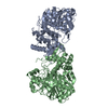
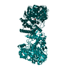
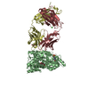
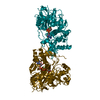
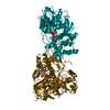
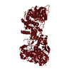
| Title | CRYSTAL STRUCTURE OF THE COMPLEX BETWEEN ESCHERICHIA COLI GLYCEROL KINASE AND THE ALLOSTERIC REGULATOR FRUCTOSE 1,6-BISPHOSPHATE. | ||||||
 Components Components | PROTEIN (GLYCEROL KINASE) | ||||||
 Keywords Keywords | TRANSFERASE / KINASE / ALLOSTERIC REGULATION / FRUCTOSE BISPHOSPHATE | ||||||
| Function / homology |  Function and homology information Function and homology informationglycerol-3-phosphate metabolic process / glycerol kinase / glycerol kinase activity / glycerol metabolic process / glycerol catabolic process / DNA damage response / zinc ion binding / ATP binding / metal ion binding / identical protein binding / cytosol Similarity search - Function | ||||||
| Biological species |  | ||||||
| Method |  X-RAY DIFFRACTION / MOLECULAR REPLACEMENT / Resolution: 3.05 Å X-RAY DIFFRACTION / MOLECULAR REPLACEMENT / Resolution: 3.05 Å | ||||||
 Authors Authors | Ormo, M. / Bystrom, C.E. / Remington, S.J. | ||||||
 Citation Citation | Journal: Biochemistry / Year: 1998 Title: Crystal structure of a complex of Escherichia coli glycerol kinase and an allosteric effector fructose 1,6-bisphosphate. Authors: Ormo, M. / Bystrom, C.E. / Remington, S.J. | ||||||
| History |
|
- Structure visualization
| Structure viewer | Molecule: MolmilJmol/JSmol |
|---|
- Downloads & links
-Download
| PDBx/mmCIF format | 1bot.cif.gz | 203.4 KB | Display | PDBx/mmCIF format |
|---|---|---|---|---|
| PDB format | pdb1bot.ent.gz | 162.8 KB | Display | PDB format |
| PDBx/mmJSON format | 1bot.json.gz | Tree view | PDBx/mmJSON format | |
| Others |  Other downloads Other downloads |
-Validation report
| Arichive directory | https://data.pdbj.org/pub/pdb/validation_reports/bo/1botftp://data.pdbj.org/pub/pdb/validation_reports/bo/1bot | HTTPS FTP |
|---|
-Related structure data
| Related structure data | 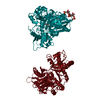 1bo5C 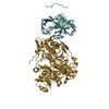 1glaS S: Starting model for refinement C: citing same article ( |
|---|---|
| Similar structure data |


-Links
 PDBj
PDBj- Assembly
| Deposited unit | 
| ||||||||
|---|---|---|---|---|---|---|---|---|---|
| 1 | 
| ||||||||
| 2 | 
| ||||||||
| Unit cell |
| ||||||||
| Noncrystallographic symmetry (NCS) | NCS oper: (Code: given Matrix: (-0.68994, -0.311658, -0.653338), Vector: |
-Components
| #1: Protein | Mass: 56162.352 Da / Num. of mol.: 2 Source method: isolated from a genetically manipulated source Source: (gene. exp.) #2: Chemical | ChemComp-EPE / |   Mass: 238.305 Da / Num. of mol.: 1 / Source method: obtained synthetically / Formula: C8H18N2O4S / Comment: pH buffer*YM Mass: 238.305 Da / Num. of mol.: 1 / Source method: obtained synthetically / Formula: C8H18N2O4S / Comment: pH buffer*YM#3: Chemical |   Mass: 92.094 Da / Num. of mol.: 2 / Source method: obtained synthetically / Formula: C3H8O3 Mass: 92.094 Da / Num. of mol.: 2 / Source method: obtained synthetically / Formula: C3H8O3 |
|---|
-Experimental details
-Experiment
| Experiment | Method: X-RAY DIFFRACTION / Number of used crystals: 1 |
|---|
- Sample preparation
| Crystal | Density Matthews: 6.4 Å3/Da / Density % sol: 81 % | |||||||||||||||||||||||||||||||||||||||||||||
|---|---|---|---|---|---|---|---|---|---|---|---|---|---|---|---|---|---|---|---|---|---|---|---|---|---|---|---|---|---|---|---|---|---|---|---|---|---|---|---|---|---|---|---|---|---|---|
| Crystal grow | pH: 7.5 Details: 1M SODIUM CITRATE, 0.1M HEPES PH 7.5, 1MM BME, CRYO PROTECTION WAS ACHIEVED BY SOAKING CRYSTAL IN MOTHER LIQUOR CONTAINING 20% GLYCEROL FOR 4 HOURS | |||||||||||||||||||||||||||||||||||||||||||||
| Crystal grow | *PLUS pH: 7 / Method: vapor diffusion, hanging drop | |||||||||||||||||||||||||||||||||||||||||||||
| Components of the solutions | *PLUS
|
-Data collection
| Diffraction | Mean temperature: 102 K |
|---|---|
| Diffraction source | Source: ROTATING ANODE / Type: RIGAKU / Wavelength: 1.5418 |
| Detector | Type: RIGAKU RAXIS II / Detector: IMAGE PLATE / Date: Mar 15, 1995 / Details: COLLIMATOR |
| Radiation | Monochromator: GRAPHITE / Protocol: SINGLE WAVELENGTH / Monochromatic (M) / Laue (L): M / Scattering type: x-ray |
| Radiation wavelength | Wavelength: 1.5418 Å / Relative weight: 1 |
| Reflection | Resolution: 3.05→20 Å / Num. obs: 45656 / % possible obs: 81 % / Observed criterion σ(I): -3 / Redundancy: 3.8 % / Biso Wilson estimate: 25.4 Å2 / Rmerge(I) obs: 0.09 |
- Processing
| Software |
| ||||||||||||||||||||||||||||||||||||||||||||||||||
|---|---|---|---|---|---|---|---|---|---|---|---|---|---|---|---|---|---|---|---|---|---|---|---|---|---|---|---|---|---|---|---|---|---|---|---|---|---|---|---|---|---|---|---|---|---|---|---|---|---|---|---|
| Refinement | Method to determine structure: MOLECULAR REPLACEMENT Starting model: 1GLA Resolution: 3.05→20 Å / Isotropic thermal model: TNT BCORREL / σ(F): 0 / Stereochemistry target values: TNT PROTGEO
| ||||||||||||||||||||||||||||||||||||||||||||||||||
| Solvent computation | Bsol: 300 Å2 / ksol: 0.75 e/Å3 | ||||||||||||||||||||||||||||||||||||||||||||||||||
| Refinement step | Cycle: LAST / Resolution: 3.05→20 Å
| ||||||||||||||||||||||||||||||||||||||||||||||||||
| Refine LS restraints |
| ||||||||||||||||||||||||||||||||||||||||||||||||||
| Software | *PLUS Name: TNT / Version: 5F / Classification: refinement | ||||||||||||||||||||||||||||||||||||||||||||||||||
| Refine LS restraints | *PLUS
|
