 Movie
Movie Controller

[English] 日本語
 Yorodumi
- PDB-1bu6: CRYSTAL STRUCTURES OF ESCHERICHIA COLI GLYCEROL KINASE AND THE MU... -
+ Open data

- Basic information
| Entry | Database: PDB / ID: 1bu6 | ||||||
|---|---|---|---|---|---|---|---|
| Title | CRYSTAL STRUCTURES OF ESCHERICHIA COLI GLYCEROL KINASE AND THE MUTANT A65T IN AN INACTIVE TETRAMER: CONFORMATIONAL CHANGES AND IMPLICATIONS FOR ALLOSTERIC REGULATION | ||||||
 Components Components | PROTEIN (GLYCEROL KINASE) | ||||||
 Keywords Keywords | TRANSFERASE / ALLOSTERY / COOPERATIVITY / GLYCEROL KINASE | ||||||
| Function / homology |  Function and homology information Function and homology informationglycerol-3-phosphate metabolic process / glycerol kinase / glycerol kinase activity / glycerol metabolic process / glycerol catabolic process / DNA damage response / zinc ion binding / ATP binding / metal ion binding / identical protein binding / cytosol Similarity search - Function | ||||||
| Biological species |  | ||||||
| Method |  X-RAY DIFFRACTION / MOLECULAR REPLACEMENT / Resolution: 2.37 Å X-RAY DIFFRACTION / MOLECULAR REPLACEMENT / Resolution: 2.37 Å | ||||||
 Authors Authors | Feese, M.D. / Faber, H.R. / Bystrom, C.E. / Pettigrew, D.W. / Remington, S.J. | ||||||
 Citation Citation | Journal: Structure / Year: 1998 Title: Glycerol kinase from Escherichia coli and an Ala65-->Thr mutant: the crystal structures reveal conformational changes with implications for allosteric regulation. Authors: Feese, M.D. / Faber, H.R. / Bystrom, C.E. / Pettigrew, D.W. / Remington, S.J. #1: Journal: J.Bacteriol. / Year: 1996Title: A Single Amino Acid Change in Escherichia Coli Glycerol Kinase Abolishes Glucose Control of Glycerol Utilization in Vivo Authors: Pettigrew, D.W. / Liu, W.Z. / Holmes, C. / Meadow, N.D. / Roseman, S. #2: Journal: Proc.Natl.Acad.Sci.USA / Year: 1994Title: Cation Promoted Association (Cpa) of a Regulatory and Target Protein is Controlled by Protein Phosphorylation Authors: Feese, M.D. / Pettigrew, D.W. / Meadow, N.D. / Roseman, S. / Remington, S.J. #3: Journal: Biochemistry / Year: 1994Title: Escherichia Coli Glycerol Kinase: Role of a Tetramer Interface in Regulation by Fructose-1,6-Bisphosphate and Phosphotransferase System Regulatory Protein Iiiglc Authors: Liu, W.Z. / Faber, H.R. / Feese, M.D. / Remington, S.J. / Pettigrew, D.W. #4: Journal: Science / Year: 1993Title: Structure of the Regulatory Complex of Escherichia Coli III==Glc== with Glycerol Kinase Authors: Hurley, J.H. / Faber, H.R. / Worthylake, D. / Meadow, N.D. / Roseman, S. / Pettigrew, D.W. / Remington, S.J. | ||||||
| History |
|
- Structure visualization
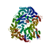
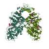
| Structure viewer | Molecule: MolmilJmol/JSmol |
|---|
- Downloads & links
-Download
| PDBx/mmCIF format | 1bu6.cif.gz | 394.6 KB | Display | PDBx/mmCIF format |
|---|---|---|---|---|
| PDB format | pdb1bu6.ent.gz | 320 KB | Display | PDB format |
| PDBx/mmJSON format | 1bu6.json.gz | Tree view | PDBx/mmJSON format | |
| Others |  Other downloads Other downloads |
-Validation report
| Arichive directory | https://data.pdbj.org/pub/pdb/validation_reports/bu/1bu6ftp://data.pdbj.org/pub/pdb/validation_reports/bu/1bu6 | HTTPS FTP |
|---|
-Related structure data
| Related structure data | 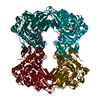 1glfSC S: Starting model for refinement C: citing same article ( |
|---|---|
| Similar structure data |


-Links
 PDBj
PDBj- Assembly
| Deposited unit | 
| ||||||||
|---|---|---|---|---|---|---|---|---|---|
| 1 |
| ||||||||
| 2 | 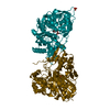
| ||||||||
| 3 | 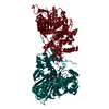
| ||||||||
| Unit cell |
|
-Components
| #1: Protein | Mass: 56192.379 Da / Num. of mol.: 4 / Mutation: A65T Source method: isolated from a genetically manipulated source Details: GLYCEROL SULPHATE / Source: (gene. exp.) #2: Chemical | ChemComp-SO4 /   Mass: 96.063 Da / Num. of mol.: 6 / Source method: obtained synthetically / Formula: SO4 Mass: 96.063 Da / Num. of mol.: 6 / Source method: obtained synthetically / Formula: SO4#3: Chemical | ChemComp-GOL /   Mass: 92.094 Da / Num. of mol.: 4 / Source method: obtained synthetically / Formula: C3H8O3 Mass: 92.094 Da / Num. of mol.: 4 / Source method: obtained synthetically / Formula: C3H8O3#4: Water | ChemComp-HOH / |  Mass: 18.015 Da / Num. of mol.: 140 / Source method: isolated from a natural source / Formula: H2O Mass: 18.015 Da / Num. of mol.: 140 / Source method: isolated from a natural source / Formula: H2O |
|---|
-Experimental details
-Experiment
| Experiment | Method: X-RAY DIFFRACTION / Number of used crystals: 1 |
|---|
- Sample preparation
| Crystal | Density Matthews: 2.65 Å3/Da / Density % sol: 53.1 % | |||||||||||||||||||||||||||||||||||||||||||||||||||||||||||||||
|---|---|---|---|---|---|---|---|---|---|---|---|---|---|---|---|---|---|---|---|---|---|---|---|---|---|---|---|---|---|---|---|---|---|---|---|---|---|---|---|---|---|---|---|---|---|---|---|---|---|---|---|---|---|---|---|---|---|---|---|---|---|---|---|---|
| Crystal grow | Method: vapor diffusion, hanging drop / pH: 8.5 Details: 20-22% (W/V) POLYETHYLENE GLYCOL MR 4000 0.2 M LISO4 0.1 M TRIS PH 8.5 - 8.8 CRYSTALLIZED BY HANGING DROP VAPOR DIFFUSION AT ROOM TEMPERATURE., vapor diffusion - hanging drop PH range: 8.5-8.8 / Temp details: room temp | |||||||||||||||||||||||||||||||||||||||||||||||||||||||||||||||
| Crystal grow | *PLUS Method: vapor diffusion, hanging drop / Details: Faber, H.R., (1989) J. Mol. Biol., 207, 637. / PH range low: 7 / PH range high: 6.5 | |||||||||||||||||||||||||||||||||||||||||||||||||||||||||||||||
| Components of the solutions | *PLUS
|
-Data collection
| Diffraction | Mean temperature: 298 K |
|---|---|
| Diffraction source | Source: ROTATING ANODE / Type: RIGAKU RU200 / Wavelength: 1.5418 |
| Detector | Type: SDMS / Detector: AREA DETECTOR / Date: Aug 15, 1994 |
| Radiation | Monochromator: GRAPHITE / Protocol: SINGLE WAVELENGTH / Monochromatic (M) / Laue (L): M / Scattering type: x-ray |
| Radiation wavelength | Wavelength: 1.5418 Å / Relative weight: 1 |
| Reflection | Resolution: 2.37→20 Å / Num. obs: 69682 / % possible obs: 74 % / Observed criterion σ(I): 2 / Redundancy: 2 % / Biso Wilson estimate: 32.3 Å2 / Rmerge(I) obs: 0.068 |
| Reflection | *PLUS Lowest resolution: 37 Å / % possible obs: 74 % / Num. measured all: 132331 |
- Processing
| Software |
| ||||||||||||||||||||||||||||||||||||||||||||||||||
|---|---|---|---|---|---|---|---|---|---|---|---|---|---|---|---|---|---|---|---|---|---|---|---|---|---|---|---|---|---|---|---|---|---|---|---|---|---|---|---|---|---|---|---|---|---|---|---|---|---|---|---|
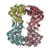
| Refinement | Method to determine structure: MOLECULAR REPLACEMENT Starting model: 1GLF Resolution: 2.37→20 Å / Isotropic thermal model: TNT BCORREL V1.0 / σ(F): 0 / Stereochemistry target values: TNT PROTGEO Details: BSOL AND KSOL SET BY USER GLYCEROL KINASE EXISTS AT PHYSIOLOGICAL CONCENTRATIONS IN AN EQUILIBRIUM BETWEEN FUNCTIONAL DIMERS AND TETRAMERS. THE CRYSTAL ASYMMETRIC UNIT CONTAINS A TETRAMER OF ...Details: BSOL AND KSOL SET BY USER GLYCEROL KINASE EXISTS AT PHYSIOLOGICAL CONCENTRATIONS IN AN EQUILIBRIUM BETWEEN FUNCTIONAL DIMERS AND TETRAMERS. THE CRYSTAL ASYMMETRIC UNIT CONTAINS A TETRAMER OF GLYCEROL KINASE WITH NEARLY EXACT 222 POINT-GROUP SYMMETRY, UNLIKE THE PREVIOUSLY SUBMITTED GLYCEROL KINASE - FACTOR IIIGLC COMPLEX IN WHICH THE 222 POINT GROUP SYMMETRY WAS CRYSTALLOGRAPHICALLY ENFORCED
| ||||||||||||||||||||||||||||||||||||||||||||||||||
| Solvent computation | Solvent model: TNT SOLVENT MODELING / Bsol: 300 Å2 / ksol: 0.8 e/Å3 | ||||||||||||||||||||||||||||||||||||||||||||||||||
| Refinement step | Cycle: LAST / Resolution: 2.37→20 Å
| ||||||||||||||||||||||||||||||||||||||||||||||||||
| Refine LS restraints |
| ||||||||||||||||||||||||||||||||||||||||||||||||||
| Software | *PLUS Name: TNT / Version: 5F-6 / Classification: refinement | ||||||||||||||||||||||||||||||||||||||||||||||||||
| Refinement | *PLUS σ(F): 0 / Rfactor obs: 0.167 | ||||||||||||||||||||||||||||||||||||||||||||||||||
| Solvent computation | *PLUS | ||||||||||||||||||||||||||||||||||||||||||||||||||
| Displacement parameters | *PLUS | ||||||||||||||||||||||||||||||||||||||||||||||||||
| Refine LS restraints | *PLUS
|
