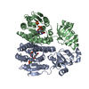
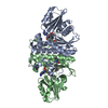
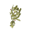
Entry Database : PDB / ID : 2w21Title Crystal structure of the aminoacid kinase domain of the glutamate 5 kinase of Escherichia coli. GLUTAMATE 5-KINASE Keywords / / / / / / / Function / homology Function Domain/homology Component
/ / / / / / / / / / / / / / / / / / / / / / / / / / / / / / / / / / / / Biological species ESCHERICHIA COLI (E. coli)Method / / / Resolution : 2.95 Å Authors Perez-Arellano, I. / Gil-Ortiz, F. / Marco-Marin, C. / Cervera, J. / Rubio, V. Journal : To be Published Title : The Structure of Glutamate 5 Kinase of Escherichia Coli without SubstratesAuthors : Perez-Arellano, I. / Gil-Ortiz, F. / Marco-Marin, C. / Cervera, J. / Rubio, V. History Deposition Oct 21, 2008 Deposition site / Processing site Revision 1.0 Nov 17, 2009 Provider / Type Revision 1.1 May 8, 2011 Group Revision 1.2 Jul 13, 2011 Group Revision 1.3 Dec 13, 2023 Group Data collection / Database references ... Data collection / Database references / Derived calculations / Other / Refinement description Category chem_comp_atom / chem_comp_bond ... chem_comp_atom / chem_comp_bond / database_2 / pdbx_database_status / pdbx_initial_refinement_model / struct_site Item _database_2.pdbx_DOI / _database_2.pdbx_database_accession ... _database_2.pdbx_DOI / _database_2.pdbx_database_accession / _pdbx_database_status.status_code_sf / _struct_site.pdbx_auth_asym_id / _struct_site.pdbx_auth_comp_id / _struct_site.pdbx_auth_seq_id
Show all Show less
 Movie
Movie Controller

 Yorodumi
 Open data
 Basic information
 Components
 Keywords
 Function and homology information
 X-RAY DIFFRACTION /
  Authors
 Citation
 Structure visualization
 Downloads & links
 Other downloads
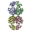


 PDBj
PDBj
 Assembly

 Mass: 96.063 Da / Num. of mol.: 4 / Source method: obtained synthetically / Formula: SO4
 Mass: 18.015 Da / Num. of mol.: 4 / Source method: isolated from a natural source / Formula: H2O
 Sample preparation
 / Beamline: BM30A / Wavelength: 0.9796
  Processing