 Movie
Movie Controller

[English] 日本語
 Yorodumi
- PDB-3lkk: Crystal structure of the isopentenyl phosphate kinase substrate c... -
+ Open data

- Basic information
| Entry | Database: PDB / ID: 3lkk | ||||||
|---|---|---|---|---|---|---|---|
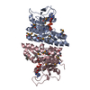
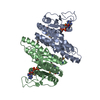
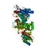
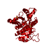
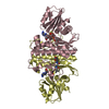
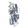
| Title | Crystal structure of the isopentenyl phosphate kinase substrate complex | ||||||
 Components Components | Gamma-glutamyl kinase related protein | ||||||
 Keywords Keywords | TRANSFERASE / isopentenyl phosphate kinase / alternate mevalonate pathway / alpha-beta-alpha sandwich fold / substrate complex / Kinase | ||||||
| Function / homology |  Function and homology information Function and homology informationisopentenyl phosphate kinase / isopentenyl phosphate kinase activity / terpenoid biosynthetic process / kinase activity / ATP binding / cytosol Similarity search - Function | ||||||
| Biological species |   Thermoplasma acidophilum (acidophilic) Thermoplasma acidophilum (acidophilic) | ||||||
| Method |  X-RAY DIFFRACTION / MOLECULAR REPLACEMENT / molecular replacement / Resolution: 2.001 Å X-RAY DIFFRACTION / MOLECULAR REPLACEMENT / molecular replacement / Resolution: 2.001 Å | ||||||
 Authors Authors | Mabanglo, M.F. / Hill, C.P. | ||||||
 Citation Citation | Journal: Acs Chem.Biol. / Year: 2010 Title: X-ray structures of isopentenyl phosphate kinase. Authors: Mabanglo, M.F. / Schubert, H.L. / Chen, M. / Hill, C.P. / Poulter, C.D. | ||||||
| History |
|
- Structure visualization
| Structure viewer | Molecule: MolmilJmol/JSmol |
|---|
- Downloads & links
-Download
| PDBx/mmCIF format | 3lkk.cif.gz | 110.6 KB | Display | PDBx/mmCIF format |
|---|---|---|---|---|
| PDB format | pdb3lkk.ent.gz | 84.1 KB | Display | PDB format |
| PDBx/mmJSON format | 3lkk.json.gz | Tree view | PDBx/mmJSON format | |
| Others |  Other downloads Other downloads |
-Validation report
| Arichive directory | https://data.pdbj.org/pub/pdb/validation_reports/lk/3lkkftp://data.pdbj.org/pub/pdb/validation_reports/lk/3lkk | HTTPS FTP |
|---|
-Related structure data


-Links
 PDBj
PDBj- Assembly
| Deposited unit | 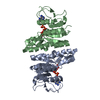
| ||||||||
|---|---|---|---|---|---|---|---|---|---|
| 1 |
| ||||||||
| Unit cell |
|
-Components
| #1: Protein | Mass: 27544.732 Da / Num. of mol.: 2 Source method: isolated from a genetically manipulated source Details: topo-cloning reaction was used to insert the Ta0103 gene, and translated protein contained histidine tag, V5 epitope and TEV protease recognition site Source: (gene. exp.) Thermoplasma acidophilum (acidophilic) / Strain: DSM1728 / Gene: Ta0103 / Plasmid: pET151/D-TOPO / Production host:  #2: Chemical |   Mass: 507.181 Da / Num. of mol.: 2 / Source method: obtained synthetically / Formula: C10H16N5O13P3 / Comment: ATP, energy-carrying molecule*YM Mass: 507.181 Da / Num. of mol.: 2 / Source method: obtained synthetically / Formula: C10H16N5O13P3 / Comment: ATP, energy-carrying molecule*YM#3: Chemical |   Mass: 166.112 Da / Num. of mol.: 2 / Source method: obtained synthetically / Formula: C5H11O4P Mass: 166.112 Da / Num. of mol.: 2 / Source method: obtained synthetically / Formula: C5H11O4P#4: Water | ChemComp-HOH / |  Mass: 18.015 Da / Num. of mol.: 198 / Source method: isolated from a natural source / Formula: H2O Mass: 18.015 Da / Num. of mol.: 198 / Source method: isolated from a natural source / Formula: H2O |
|---|
-Experimental details
-Experiment
| Experiment | Method: X-RAY DIFFRACTION / Number of used crystals: 1 |
|---|
- Sample preparation
| Crystal | Density Matthews: 2.18 Å3/Da / Density % sol: 43.53 % |
|---|---|
| Crystal grow | Temperature: 274 K / Method: vapor diffusion, sitting drop / pH: 7 Details: 0.1 M MIB buffer (2:3:3 molar ratio of sodium malonate, imidazole, boric acid), pH 7.0, 25% PEG1500, VAPOR DIFFUSION, SITTING DROP, temperature 274K |
-Data collection
| Diffraction | Mean temperature: 100 K |
|---|---|
| Diffraction source | Source: ROTATING ANODE / Type: RIGAKU MICROMAX-007 HF / Wavelength: 1.5418 Å |
| Detector | Type: RIGAKU RAXIS IV / Detector: IMAGE PLATE / Date: Sep 15, 2008 / Details: mirrors |
| Radiation | Monochromator: Verimax / Protocol: SINGLE WAVELENGTH / Monochromatic (M) / Laue (L): M / Scattering type: x-ray |
| Radiation wavelength | Wavelength: 1.5418 Å / Relative weight: 1 |
| Reflection | Resolution: 2→30 Å / Num. obs: 31502 / % possible obs: 98 % / Redundancy: 3.7 % / Rmerge(I) obs: 0.075 / Net I/σ(I): 11.2 |
| Reflection shell | Resolution: 2→2.07 Å / Redundancy: 3.3 % / Rmerge(I) obs: 0.475 / % possible all: 95.2 |
-Phasing
| Phasing | Method: molecular replacement | |||||||||
|---|---|---|---|---|---|---|---|---|---|---|
| Phasing MR | Model details: Phaser MODE: MR_AUTO
|
- Processing
| Software |
| ||||||||||||||||||||||||||||||||||||||||||||||||||||||||||||||||||||||||||||||||||||
|---|---|---|---|---|---|---|---|---|---|---|---|---|---|---|---|---|---|---|---|---|---|---|---|---|---|---|---|---|---|---|---|---|---|---|---|---|---|---|---|---|---|---|---|---|---|---|---|---|---|---|---|---|---|---|---|---|---|---|---|---|---|---|---|---|---|---|---|---|---|---|---|---|---|---|---|---|---|---|---|---|---|---|---|---|---|
| Refinement | Method to determine structure: MOLECULAR REPLACEMENT Starting model: T.acidophilum isopentenyl phosphate kinase solved by single-wavelength anomalous diffraction (SAD) Resolution: 2.001→29.303 Å / Occupancy max: 1 / Occupancy min: 0 / SU ML: 0.28 / Isotropic thermal model: Isotropic / σ(F): 1.34 / Phase error: 22.47 / Stereochemistry target values: ML
| ||||||||||||||||||||||||||||||||||||||||||||||||||||||||||||||||||||||||||||||||||||
| Solvent computation | Shrinkage radii: 0.9 Å / VDW probe radii: 1.11 Å / Solvent model: FLAT BULK SOLVENT MODEL / Bsol: 40.151 Å2 / ksol: 0.4 e/Å3 | ||||||||||||||||||||||||||||||||||||||||||||||||||||||||||||||||||||||||||||||||||||
| Displacement parameters |
| ||||||||||||||||||||||||||||||||||||||||||||||||||||||||||||||||||||||||||||||||||||
| Refinement step | Cycle: LAST / Resolution: 2.001→29.303 Å
| ||||||||||||||||||||||||||||||||||||||||||||||||||||||||||||||||||||||||||||||||||||
| Refine LS restraints |
| ||||||||||||||||||||||||||||||||||||||||||||||||||||||||||||||||||||||||||||||||||||
| LS refinement shell |
|
