 Movie
Movie Controller

[English] 日本語
 Yorodumi
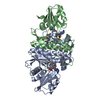
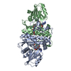
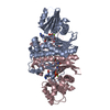
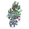
- PDB-6awh: Staphylococcus aureus Type II pantothenate kinase in complex with... -
+ Open data

- Basic information
| Entry | Database: PDB / ID: 6awh | ||||||
|---|---|---|---|---|---|---|---|
| Title | Staphylococcus aureus Type II pantothenate kinase in complex with ATP and pantothenate analog Deoxy-MeO-N5Pan | ||||||
 Components Components | Type II pantothenate kinase | ||||||
 Keywords Keywords | TRANSFERASE / Staphylococcus aureus / SaPanK / ATP / pantothenate analog | ||||||
| Function / homology |  Function and homology information Function and homology informationpantothenate kinase / pantothenate kinase activity / coenzyme A biosynthetic process / ATP binding / cytosol Similarity search - Function | ||||||
| Biological species |   Staphylococcus aureus (bacteria) Staphylococcus aureus (bacteria) | ||||||
| Method |  X-RAY DIFFRACTION / SYNCHROTRON / MOLECULAR REPLACEMENT / Resolution: 1.9 Å X-RAY DIFFRACTION / SYNCHROTRON / MOLECULAR REPLACEMENT / Resolution: 1.9 Å | ||||||
 Authors Authors | Chen, Y. / Antoshchenko, T. / Strauss, E. / Barnard, L. / Huang, Y.H. | ||||||
 Citation Citation | Journal: To Be Published Title: Structure-based identification of uncompetitive inhibitors for Staphylococcus aureus pantothenate kinase. Authors: Chen, Y. / Antoshchenko, T. / Strauss, E. / Barnard, L. / Huang, Y.H. | ||||||
| History |
|
- Structure visualization
| Structure viewer | Molecule: MolmilJmol/JSmol |
|---|
- Downloads & links
-Download
| PDBx/mmCIF format | 6awh.cif.gz | 231.3 KB | Display | PDBx/mmCIF format |
|---|---|---|---|---|
| PDB format | pdb6awh.ent.gz | 184.7 KB | Display | PDB format |
| PDBx/mmJSON format | 6awh.json.gz | Tree view | PDBx/mmJSON format | |
| Others |  Other downloads Other downloads |
-Validation report
| Arichive directory | https://data.pdbj.org/pub/pdb/validation_reports/aw/6awhftp://data.pdbj.org/pub/pdb/validation_reports/aw/6awh | HTTPS FTP |
|---|
-Related structure data


-Links
 PDBj
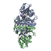
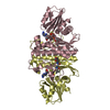
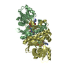
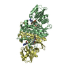
PDBj- Assembly
| Deposited unit | 
| ||||||||
|---|---|---|---|---|---|---|---|---|---|
| 1 | 
| ||||||||
| 2 | 
| ||||||||
| Unit cell |
|
-Components
| #1: Protein | Mass: 28997.770 Da / Num. of mol.: 4 Source method: isolated from a genetically manipulated source Source: (gene. exp.) Staphylococcus aureus (bacteria)Gene: coaW, AYM28_11860, AYM37_11860, ERS072738_01917, ERS074020_01878, HMPREF3211_02599 Production host: References: UniProt: A0A0D6HHM8, UniProt: Q2FWC7*PLUS, pantothenate kinase #2: Chemical | ChemComp-ATP /   Mass: 507.181 Da / Num. of mol.: 4 / Source method: obtained synthetically / Formula: C10H16N5O13P3 / Comment: ATP, energy-carrying molecule*YM Mass: 507.181 Da / Num. of mol.: 4 / Source method: obtained synthetically / Formula: C10H16N5O13P3 / Comment: ATP, energy-carrying molecule*YM#3: Chemical | ChemComp-MG /   Mass: 24.305 Da / Num. of mol.: 4 / Source method: obtained synthetically / Formula: Mg Mass: 24.305 Da / Num. of mol.: 4 / Source method: obtained synthetically / Formula: Mg#4: Chemical | ChemComp-N7G / (   Mass: 302.410 Da / Num. of mol.: 4 / Source method: obtained synthetically / Formula: C15H30N2O4 / Feature type: SUBJECT OF INVESTIGATION Mass: 302.410 Da / Num. of mol.: 4 / Source method: obtained synthetically / Formula: C15H30N2O4 / Feature type: SUBJECT OF INVESTIGATION#5: Water | ChemComp-HOH / |  Mass: 18.015 Da / Num. of mol.: 635 / Source method: isolated from a natural source / Formula: H2O Mass: 18.015 Da / Num. of mol.: 635 / Source method: isolated from a natural source / Formula: H2O |
|---|
-Experimental details
-Experiment
| Experiment | Method: X-RAY DIFFRACTION / Number of used crystals: 1 |
|---|
- Sample preparation
| Crystal | Density Matthews: 2.36 Å3/Da / Density % sol: 47.99 % |
|---|---|
| Crystal grow | Temperature: 289.15 K / Method: vapor diffusion, sitting drop / pH: 6.5 Details: 23% PEG3350, 0.1 M ammonium sulfate, 0.1 M Bis-Tris, pH 6.5 |
-Data collection
| Diffraction | Mean temperature: 100 K |
|---|---|
| Diffraction source | Source: SYNCHROTRON / Site: CLSI  / Beamline: 08ID-1 / Wavelength: 0.9795 Å / Beamline: 08ID-1 / Wavelength: 0.9795 Å |
| Detector | Type: RAYONIX MX-300 / Detector: CCD / Date: Dec 17, 2016 |
| Radiation | Monochromator: double crystal Si(111) / Protocol: SINGLE WAVELENGTH / Monochromatic (M) / Laue (L): M / Scattering type: x-ray |
| Radiation wavelength | Wavelength: 0.9795 Å / Relative weight: 1 |
| Reflection | Resolution: 1.9→98.27 Å / Num. obs: 87345 / % possible obs: 99.4 % / Redundancy: 5.56 % / Net I/σ(I): 4.9 |
| Reflection shell | Highest resolution: 1.9 Å |
- Processing
| Software |
| ||||||||||||||||||||||||||||||||||||||||||||||||||||||||||||
|---|---|---|---|---|---|---|---|---|---|---|---|---|---|---|---|---|---|---|---|---|---|---|---|---|---|---|---|---|---|---|---|---|---|---|---|---|---|---|---|---|---|---|---|---|---|---|---|---|---|---|---|---|---|---|---|---|---|---|---|---|---|
| Refinement | Method to determine structure: MOLECULAR REPLACEMENT / Resolution: 1.9→98.27 Å / Cor.coef. Fo:Fc: 0.962 / Cor.coef. Fo:Fc free: 0.952 / SU B: 4.494 / SU ML: 0.123 / Cross valid method: THROUGHOUT / σ(F): 0 / ESU R: 0.165 / ESU R Free: 0.144 Details: HYDROGENS HAVE BEEN ADDED IN THE RIDING POSITIONS U VALUES : REFINED INDIVIDUALLY
| ||||||||||||||||||||||||||||||||||||||||||||||||||||||||||||
| Solvent computation | Ion probe radii: 0.8 Å / Shrinkage radii: 0.8 Å / VDW probe radii: 1.2 Å | ||||||||||||||||||||||||||||||||||||||||||||||||||||||||||||
| Displacement parameters | Biso max: 104.71 Å2 / Biso mean: 29.326 Å2 / Biso min: 12.92 Å2
| ||||||||||||||||||||||||||||||||||||||||||||||||||||||||||||
| Refinement step | Cycle: final / Resolution: 1.9→98.27 Å
| ||||||||||||||||||||||||||||||||||||||||||||||||||||||||||||
| Refine LS restraints |
| ||||||||||||||||||||||||||||||||||||||||||||||||||||||||||||
| LS refinement shell | Resolution: 1.898→1.947 Å / Rfactor Rfree error: 0 / Total num. of bins used: 20
|
