 Movie
Movie Controller

[English] 日本語
 Yorodumi
- PDB-2vz0: Pteridine Reductase 1 (PTR1) from Trypanosoma Brucei in complex w... -
+ Open data

- Basic information
| Entry | Database: PDB / ID: 2vz0 | ||||||
|---|---|---|---|---|---|---|---|
| Title | Pteridine Reductase 1 (PTR1) from Trypanosoma Brucei in complex with NADP and DDD00066641 | ||||||
 Components Components | PTERIDINE REDUCTASE | ||||||
 Keywords Keywords | OXIDOREDUCTASE / SHORT-CHAIN DEHYDROGENASE/REDUCTASE / TRYPANOSOMATIDS / PTERIDINE REDUCTASE | ||||||
| Function / homology |  Function and homology information Function and homology information | ||||||
| Biological species |  | ||||||
| Method |  X-RAY DIFFRACTION / MOLECULAR REPLACEMENT / Resolution: 1.9 Å X-RAY DIFFRACTION / MOLECULAR REPLACEMENT / Resolution: 1.9 Å | ||||||
 Authors Authors | Robinson, D.A. / Thompson, S. / Sienkiewicz, N. / Fairlamb, A.H. | ||||||
 Citation Citation | Journal: Anal.Biochem. / Year: 2010 Title: Development and Validation of a Cytochrome C Coupled Assay for Pteridine Reductase 1 and Dihydrofolate Reductase. Authors: Shanks, E.J. / Ong, H.B. / Robinson, D.A. / Thompson, S. / Sienkiewicz, N. / Fairlamb, A.H. / Frearson, J.A. #1: Journal: Mol.Microbiol. / Year: 2006Title: Structure and Reactivity of Trypanosoma Brucei Pteridine Reductase: Inhibition by the Archetypal Antifolate Methotrexate. Authors: Dawson, A. / Gibellini, F. / Sienkiewicz, N. / Tulloch, L.B. / Fyfe, P.K. / Mcluskey, K. / Fairlamb, A.H. / Hunter, W.N. | ||||||
| History |
|
- Structure visualization
| Structure viewer | Molecule: MolmilJmol/JSmol |
|---|
- Downloads & links
-Download
| PDBx/mmCIF format | 2vz0.cif.gz | 224 KB | Display | PDBx/mmCIF format |
|---|---|---|---|---|
| PDB format | pdb2vz0.ent.gz | 180.3 KB | Display | PDB format |
| PDBx/mmJSON format | 2vz0.json.gz | Tree view | PDBx/mmJSON format | |
| Others |  Other downloads Other downloads |
-Validation report
| Arichive directory | https://data.pdbj.org/pub/pdb/validation_reports/vz/2vz0ftp://data.pdbj.org/pub/pdb/validation_reports/vz/2vz0 | HTTPS FTP |
|---|
-Related structure data
| Related structure data |  2c7vS S: Starting model for refinement |
|---|---|
| Similar structure data |


-Links
 PDBj
PDBj


- Assembly
| Deposited unit | 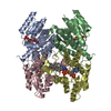
| ||||||||||||||||
|---|---|---|---|---|---|---|---|---|---|---|---|---|---|---|---|---|---|
| 1 |
| ||||||||||||||||
| Unit cell |
| ||||||||||||||||
| Noncrystallographic symmetry (NCS) | NCS oper:
|
-Components
| #1: Protein | Mass: 28498.441 Da / Num. of mol.: 4 Source method: isolated from a genetically manipulated source Source: (gene. exp.)  #2: Chemical | ChemComp-NAP /   Mass: 743.405 Da / Num. of mol.: 4 / Source method: obtained synthetically / Formula: C21H28N7O17P3 Mass: 743.405 Da / Num. of mol.: 4 / Source method: obtained synthetically / Formula: C21H28N7O17P3#3: Chemical | ChemComp-D64 /   Mass: 250.298 Da / Num. of mol.: 4 / Source method: obtained synthetically / Formula: C15H14N4 Mass: 250.298 Da / Num. of mol.: 4 / Source method: obtained synthetically / Formula: C15H14N4#4: Water | ChemComp-HOH / |  Mass: 18.015 Da / Num. of mol.: 1146 / Source method: isolated from a natural source / Formula: H2O Mass: 18.015 Da / Num. of mol.: 1146 / Source method: isolated from a natural source / Formula: H2ONonpolymer details | 6-P-TOLYL-QUINAZOLIN | |
|---|
-Experimental details
-Experiment
| Experiment | Method: X-RAY DIFFRACTION / Number of used crystals: 1 |
|---|
- Sample preparation
| Crystal | Density Matthews: 1.9 Å3/Da / Density % sol: 38 % / Description: NONE |
|---|---|
| Crystal grow | pH: 6 / Details: 1.8M NA ACETATE, 0.1M CITRATE BUFFER PH6.0 |
-Data collection
| Diffraction | Mean temperature: 100 K |
|---|---|
| Diffraction source | Source: ROTATING ANODE / Type: RIGAKU MICROMAX-007 / Wavelength: 1.5418 |
| Detector | Type: RIGAKU IMAGE PLATE / Detector: IMAGE PLATE / Date: Jul 17, 2008 / Details: MIRRORS |
| Radiation | Protocol: SINGLE WAVELENGTH / Monochromatic (M) / Laue (L): M / Scattering type: x-ray |
| Radiation wavelength | Wavelength: 1.5418 Å / Relative weight: 1 |
| Reflection | Resolution: 1.9→20 Å / Num. obs: 72743 / % possible obs: 93 % / Observed criterion σ(I): 0 / Redundancy: 2.4 % / Rmerge(I) obs: 0.04 / Net I/σ(I): 16 |
| Reflection shell | Resolution: 1.9→2 Å / Redundancy: 1.7 % / Rmerge(I) obs: 0.39 / Mean I/σ(I) obs: 3 / % possible all: 75.1 |
- Processing
| Software |
| ||||||||||||||||||||||||||||||||||||||||||||||||||||||||||||||||||||||||||||||||||||||||||||||||||||||||||||||||||||||||||||||||||||||||||||||||||||||||||||||||||||||||||||||||||||||
|---|---|---|---|---|---|---|---|---|---|---|---|---|---|---|---|---|---|---|---|---|---|---|---|---|---|---|---|---|---|---|---|---|---|---|---|---|---|---|---|---|---|---|---|---|---|---|---|---|---|---|---|---|---|---|---|---|---|---|---|---|---|---|---|---|---|---|---|---|---|---|---|---|---|---|---|---|---|---|---|---|---|---|---|---|---|---|---|---|---|---|---|---|---|---|---|---|---|---|---|---|---|---|---|---|---|---|---|---|---|---|---|---|---|---|---|---|---|---|---|---|---|---|---|---|---|---|---|---|---|---|---|---|---|---|---|---|---|---|---|---|---|---|---|---|---|---|---|---|---|---|---|---|---|---|---|---|---|---|---|---|---|---|---|---|---|---|---|---|---|---|---|---|---|---|---|---|---|---|---|---|---|---|---|
| Refinement | Method to determine structure: MOLECULAR REPLACEMENT Starting model: PDB ENTRY 2C7V Resolution: 1.9→20 Å / Cor.coef. Fo:Fc: 0.966 / Cor.coef. Fo:Fc free: 0.94 / SU B: 5.662 / SU ML: 0.093 / TLS residual ADP flag: LIKELY RESIDUAL / Cross valid method: THROUGHOUT / ESU R: 0.156 / ESU R Free: 0.144 / Stereochemistry target values: MAXIMUM LIKELIHOOD / Details: HYDROGENS HAVE BEEN ADDED IN THE RIDING POSITIONS.
| ||||||||||||||||||||||||||||||||||||||||||||||||||||||||||||||||||||||||||||||||||||||||||||||||||||||||||||||||||||||||||||||||||||||||||||||||||||||||||||||||||||||||||||||||||||||
| Solvent computation | Ion probe radii: 0.8 Å / Shrinkage radii: 0.8 Å / VDW probe radii: 1.2 Å / Solvent model: MASK | ||||||||||||||||||||||||||||||||||||||||||||||||||||||||||||||||||||||||||||||||||||||||||||||||||||||||||||||||||||||||||||||||||||||||||||||||||||||||||||||||||||||||||||||||||||||
| Displacement parameters | Biso mean: 22.64 Å2
| ||||||||||||||||||||||||||||||||||||||||||||||||||||||||||||||||||||||||||||||||||||||||||||||||||||||||||||||||||||||||||||||||||||||||||||||||||||||||||||||||||||||||||||||||||||||
| Refinement step | Cycle: LAST / Resolution: 1.9→20 Å
| ||||||||||||||||||||||||||||||||||||||||||||||||||||||||||||||||||||||||||||||||||||||||||||||||||||||||||||||||||||||||||||||||||||||||||||||||||||||||||||||||||||||||||||||||||||||
| Refine LS restraints |
|