 Movie
Movie Controller

[English] 日本語
 Yorodumi
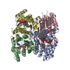
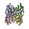
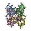
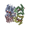
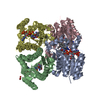
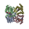
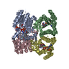
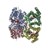
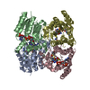
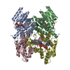
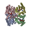
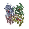
- PDB-4cm4: Crystal structure of pteridine reductase 1 (PTR1) from Trypanosom... -
+ Open data

- Basic information
| Entry | Database: PDB / ID: 4cm4 | ||||||
|---|---|---|---|---|---|---|---|
| Title | Crystal structure of pteridine reductase 1 (PTR1) from Trypanosoma brucei in ternary complex with cofactor and inhibitor | ||||||
 Components Components | (PTERIDINE REDUCTASE ...) x 2 | ||||||
 Keywords Keywords | OXIDOREDUCTASE / PTR1 / SHORT-CHAIN DEHYDROGENASE/REDUCTASE | ||||||
| Function / homology |  Function and homology information Function and homology information | ||||||
| Biological species |  | ||||||
| Method |  X-RAY DIFFRACTION / OTHER / Resolution: 1.81 Å X-RAY DIFFRACTION / OTHER / Resolution: 1.81 Å | ||||||
 Authors Authors | Barrack, K.L. / Hunter, W.N. | ||||||
 Citation Citation | Journal: J.Med.Chem. / Year: 2014 Title: Structure-Based Design and Synthesis of Antiparasitic Pyrrolopyrimidines Targeting Pteridine Reductase 1. Authors: Khalaf, A.I. / Huggan, J.K. / Suckling, C.J. / Gibson, C.L. / Stewart, K. / Giordani, F. / Barrett, M.P. / Wong, P.E. / Barrack, K.L. / Hunter, W.N. | ||||||
| History |
|
- Structure visualization
| Structure viewer | Molecule: MolmilJmol/JSmol |
|---|
- Downloads & links
-Download
| PDBx/mmCIF format | 4cm4.cif.gz | 220.8 KB | Display | PDBx/mmCIF format |
|---|---|---|---|---|
| PDB format | pdb4cm4.ent.gz | 175.7 KB | Display | PDB format |
| PDBx/mmJSON format | 4cm4.json.gz | Tree view | PDBx/mmJSON format | |
| Others |  Other downloads Other downloads |
-Validation report
| Arichive directory | https://data.pdbj.org/pub/pdb/validation_reports/cm/4cm4ftp://data.pdbj.org/pub/pdb/validation_reports/cm/4cm4 | HTTPS FTP |
|---|
-Related structure data
| Related structure data |  4cl8C 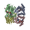 4cldC 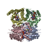 4cleC 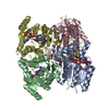 4clhC 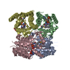 4cloC 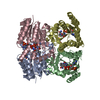 4clrC  4clxC  4cm1C  4cm3C  4cm5C  4cm6C  4cm7C  4cm8C  4cm9C  4cmaC  4cmbC 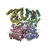 4cmcC 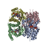 4cmeC  4cmgC  4cmiC 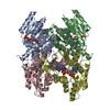 4cmjC  4cmkC C: citing same article ( |
|---|---|
| Similar structure data |


-Links
 PDBj
PDBj


- Assembly
| Deposited unit | 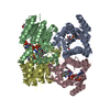
| ||||||||||||||||
|---|---|---|---|---|---|---|---|---|---|---|---|---|---|---|---|---|---|
| 1 |
| ||||||||||||||||
| Unit cell |
| ||||||||||||||||
| Noncrystallographic symmetry (NCS) | NCS oper:
|
-Components
-PTERIDINE REDUCTASE ... , 2 types, 4 molecules ABCD
| #1: Protein | Mass: 30685.787 Da / Num. of mol.: 1 Source method: isolated from a genetically manipulated source Source: (gene. exp.)  |
|---|---|
| #2: Protein | Mass: 30669.791 Da / Num. of mol.: 3 Source method: isolated from a genetically manipulated source Source: (gene. exp.) |
-Non-polymers , 5 types, 729 molecules 
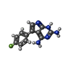


| #3: Chemical | ChemComp-NAP /  Mass: 743.405 Da / Num. of mol.: 4 / Source method: obtained synthetically / Formula: C21H28N7O17P3 Mass: 743.405 Da / Num. of mol.: 4 / Source method: obtained synthetically / Formula: C21H28N7O17P3#4: Chemical | ChemComp-4NR /  Mass: 243.240 Da / Num. of mol.: 4 / Source method: obtained synthetically / Formula: C12H10FN5 Mass: 243.240 Da / Num. of mol.: 4 / Source method: obtained synthetically / Formula: C12H10FN5#5: Chemical | ChemComp-GOL / |  Mass: 92.094 Da / Num. of mol.: 1 / Source method: obtained synthetically / Formula: C3H8O3 Mass: 92.094 Da / Num. of mol.: 1 / Source method: obtained synthetically / Formula: C3H8O3#6: Chemical |  Mass: 59.044 Da / Num. of mol.: 2 / Source method: obtained synthetically / Formula: C2H3O2 Mass: 59.044 Da / Num. of mol.: 2 / Source method: obtained synthetically / Formula: C2H3O2#7: Water | ChemComp-HOH / | Mass: 18.015 Da / Num. of mol.: 718 / Source method: isolated from a natural source / Formula: H2O |
|---|
-Details
| Has protein modification | Y |
|---|---|
| Nonpolymer details | S-OXY CYSTEINE (CSX): CYSTEINE 168 MODIFIED TO S-OXY CYSTEINE IN CHAIN A |
| Sequence details | SEQUENCE CONTAINS ADDITIONAL |
-Experimental details
-Experiment
| Experiment | Method: X-RAY DIFFRACTION / Number of used crystals: 1 |
|---|
- Sample preparation
| Crystal | Density Matthews: 2.04 Å3/Da / Density % sol: 39.57 % / Description: NONE |
|---|---|
| Crystal grow | Details: RESERVOIR CONTAINED 1.7-2.7 M SODIUM ACETATE, 20-50 MM SODIUM CITRATE PH 4.5-5.0 |
-Data collection
| Diffraction | Mean temperature: 100 K |
|---|---|
| Diffraction source | Source: ROTATING ANODE / Type: RIGAKU MICROMAX-007 HF / Wavelength: 1.5418 |
| Detector | Type: RIGAKU R-AXIS IV / Detector: IMAGE PLATE / Date: Sep 22, 2011 |
| Radiation | Protocol: SINGLE WAVELENGTH / Monochromatic (M) / Laue (L): M / Scattering type: x-ray |
| Radiation wavelength | Wavelength: 1.5418 Å / Relative weight: 1 |
| Reflection | Resolution: 1.81→19.76 Å / Num. obs: 83224 / % possible obs: 92.6 % / Redundancy: 5.1 % / Rmerge(I) obs: 0.04 / Net I/σ(I): 24.7 |
| Reflection shell | Resolution: 1.81→1.9 Å / Redundancy: 4.5 % / Rmerge(I) obs: 0.19 / Mean I/σ(I) obs: 8.2 / % possible all: 77.1 |
- Processing
| Software |
| ||||||||||||||||||||||||||||||||||||||||||||||||||||||||||||||||||||||||||||||||||||||||||||||||||||||||||||||||||||||||||||||||||||||||||||||||||||||||||||||||||||||||||||||||||||||
|---|---|---|---|---|---|---|---|---|---|---|---|---|---|---|---|---|---|---|---|---|---|---|---|---|---|---|---|---|---|---|---|---|---|---|---|---|---|---|---|---|---|---|---|---|---|---|---|---|---|---|---|---|---|---|---|---|---|---|---|---|---|---|---|---|---|---|---|---|---|---|---|---|---|---|---|---|---|---|---|---|---|---|---|---|---|---|---|---|---|---|---|---|---|---|---|---|---|---|---|---|---|---|---|---|---|---|---|---|---|---|---|---|---|---|---|---|---|---|---|---|---|---|---|---|---|---|---|---|---|---|---|---|---|---|---|---|---|---|---|---|---|---|---|---|---|---|---|---|---|---|---|---|---|---|---|---|---|---|---|---|---|---|---|---|---|---|---|---|---|---|---|---|---|---|---|---|---|---|---|---|---|---|---|
| Refinement | Method to determine structure: OTHER Starting model: NONE Resolution: 1.81→19.3 Å / Cor.coef. Fo:Fc: 0.971 / Cor.coef. Fo:Fc free: 0.958 / SU B: 2.2 / SU ML: 0.069 / Cross valid method: THROUGHOUT / ESU R: 0.12 / ESU R Free: 0.113 / Stereochemistry target values: MAXIMUM LIKELIHOOD Details: HYDROGENS HAVE BEEN ADDED IN THE RIDING POSITIONS. RESIDUES WITH INSUFFICIENT ELECTRON DENSITY WERE NOT MODELED.
| ||||||||||||||||||||||||||||||||||||||||||||||||||||||||||||||||||||||||||||||||||||||||||||||||||||||||||||||||||||||||||||||||||||||||||||||||||||||||||||||||||||||||||||||||||||||
| Solvent computation | Ion probe radii: 0.8 Å / Shrinkage radii: 0.8 Å / VDW probe radii: 1.2 Å / Solvent model: MASK | ||||||||||||||||||||||||||||||||||||||||||||||||||||||||||||||||||||||||||||||||||||||||||||||||||||||||||||||||||||||||||||||||||||||||||||||||||||||||||||||||||||||||||||||||||||||
| Displacement parameters | Biso mean: 19.061 Å2
| ||||||||||||||||||||||||||||||||||||||||||||||||||||||||||||||||||||||||||||||||||||||||||||||||||||||||||||||||||||||||||||||||||||||||||||||||||||||||||||||||||||||||||||||||||||||
| Refinement step | Cycle: LAST / Resolution: 1.81→19.3 Å
| ||||||||||||||||||||||||||||||||||||||||||||||||||||||||||||||||||||||||||||||||||||||||||||||||||||||||||||||||||||||||||||||||||||||||||||||||||||||||||||||||||||||||||||||||||||||
| Refine LS restraints |
|