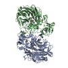
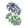
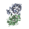
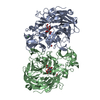
[English] 日本語
Yorodumi
- PDB-2vvz: Structure of the catalytic domain of Streptococcus pneumoniae sia... -

+
Open data


ID or keywords:

Loading...

-
Basic information

Entry
Database: PDB / ID: 2vvz
TitleStructure of the catalytic domain of Streptococcus pneumoniae sialidase NanA
ComponentsSIALIDASE A
KeywordsHYDROLASE / SECRETED / CELL WALL / SIALIDASE / PEPTIDOGLYCAN-ANCHOR / GLYCOSIDASE / NEURAMINIDASE / VIRULENCE FACTOR
Function / homology
Function and homology information


ganglioside catabolic process / oligosaccharide catabolic process / exo-alpha-(2->3)-sialidase activity / exo-alpha-(2->6)-sialidase activity / exo-alpha-(2->8)-sialidase activity / exo-alpha-sialidase / intracellular membrane-bounded organelle / extracellular region / membrane / cytoplasm
Similarity search - Function
Intramolecular trans-sialidase; domain 3 / Intramolecular Trans-sialidase; Domain 3 / Trans-sialidase, domain 3 / Glycoside hydrolase, family 33, N-terminal / Sialidase, N-terminal domain / BNR repeat-like domain / Laminin G domain / Laminin G domain / Sialidase family / Sialidase ...Intramolecular trans-sialidase; domain 3 / Intramolecular Trans-sialidase; Domain 3 / Trans-sialidase, domain 3 / Glycoside hydrolase, family 33, N-terminal / Sialidase, N-terminal domain / BNR repeat-like domain / Laminin G domain / Laminin G domain / Sialidase family / Sialidase / YSIRK type signal peptide / Neuraminidase - #10 / YSIRK Gram-positive signal peptide / Gram-positive cocci surface proteins LPxTG motif profile. / Sialidase superfamily / LPXTG cell wall anchor domain / 6 Propeller / Neuraminidase / Concanavalin A-like lectin/glucanase domain superfamily / Beta Barrel / Mainly Beta
Similarity search - Domain/homology
2-DEOXY-2,3-DEHYDRO-N-ACETYL-NEURAMINIC ACID / Sialidase A
Similarity search - Component
Biological speciesSTREPTOCOCCUS PNEUMONIAE (bacteria)
MethodX-RAY DIFFRACTION / MOLECULAR REPLACEMENT / Resolution: 2.5 Å
AuthorsXu, G. / Li, X. / Andrew, P.W. / Taylor, G.L.
CitationJournal: Acta Crystallogr.,Sect.F / Year: 2008
Title: Structure of the Catalytic Domain of Streptococcus Pneumoniae Sialidase Nana.
Authors: Xu, G. / Li, X. / Andrew, P.W. / Taylor, G.L.
History
DepositionJun 13, 2008Deposition site: PDBE / Processing site: PDBE
Revision 1.0Jun 24, 2008Provider: repository / Type: Initial release
Revision 1.1May 8, 2011Group: Version format compliance
Revision 1.2Jul 13, 2011Group: Version format compliance
Revision 1.3Jul 5, 2017Group: Data collection / Category: diffrn_source / Item: _diffrn_source.type
Revision 1.4Jul 29, 2020Group: Data collection / Derived calculations / Other
Category: chem_comp / pdbx_database_status ...chem_comp / pdbx_database_status / struct_site / struct_site_gen
Item: _chem_comp.type / _pdbx_database_status.status_code_sf
Description: Carbohydrate remediation / Provider: repository / Type: Remediation
Revision 1.5Sep 2, 2020Group: Derived calculations / Structure summary
Category: chem_comp / pdbx_struct_assembly ...chem_comp / pdbx_struct_assembly / pdbx_struct_assembly_gen / pdbx_struct_assembly_prop
Item: _chem_comp.pdbx_synonyms
Revision 1.6Dec 13, 2023Group: Data collection / Database references / Refinement description
Category: chem_comp_atom / chem_comp_bond ...chem_comp_atom / chem_comp_bond / database_2 / pdbx_initial_refinement_model
Item: _database_2.pdbx_DOI / _database_2.pdbx_database_accession
Remark 700 SHEET THE SHEET STRUCTURE OF THIS MOLECULE IS BIFURCATED. IN ORDER TO REPRESENT THIS FEATURE IN ... SHEET THE SHEET STRUCTURE OF THIS MOLECULE IS BIFURCATED. IN ORDER TO REPRESENT THIS FEATURE IN THE SHEET RECORDS BELOW, TWO SHEETS ARE DEFINED.

-
Structure visualization

Structure viewerMolecule:
MolmilJmol/JSmol

Downloads & links

-
Assembly

Deposited unit
A: SIALIDASE A
B: SIALIDASE A
hetero molecules


Theoretical massNumber of molelcules
Total (without water)113,6955
Polymers113,0772
Non-polymers6183
Water2,234124
1


  • Idetical with deposited unit
  • defined by software
TypeNameSymmetry operationNumber
identity operation1_555x,y,z1
Buried area3790 Å2
ΔGint-13 kcal/mol
Surface area35820 Å2
MethodPISA
Unit cell
Length a, b, c (Å)49.206, 95.626, 226.596
Angle α, β, γ (deg.)90.00, 90.00, 90.00
Int Tables number19
Space group name H-MP212121

-
Components

#1: Protein SIALIDASE A / NANA SIALIDASE / NEURAMINIDASE A


Mass: 56538.332 Da / Num. of mol.: 2 / Fragment: CATALYTIC DOMAIN, RESIDUES 319-822
Source method: isolated from a genetically manipulated source
Source: (gene. exp.) STREPTOCOCCUS PNEUMONIAE (bacteria) / Strain: R36A / Production host: ESCHERICHIA COLI (E. coli) / Strain (production host): BL21(DE3) / References: UniProt: P62575, exo-alpha-sialidase
#2: Sugar ChemComp-DAN / 2-DEOXY-2,3-DEHYDRO-N-ACETYL-NEURAMINIC ACID / Neu5Ac2en


Type: D-saccharide / Mass: 291.255 Da / Num. of mol.: 2 / Source method: obtained synthetically / Formula: C11H17NO8
#3: Chemical ChemComp-CL / CHLORIDE ION


Mass: 35.453 Da / Num. of mol.: 1 / Source method: obtained synthetically / Formula: Cl
#4: Water ChemComp-HOH / water


Mass: 18.015 Da / Num. of mol.: 124 / Source method: isolated from a natural source / Formula: H2O

-
Experimental details

-
Experiment

ExperimentMethod: X-RAY DIFFRACTION / Number of used crystals: 1

-
Sample preparation

CrystalDensity Matthews: 2.36 Å3/Da / Density % sol: 48 % / Description: NONE
Crystal growpH: 6.4 / Details: 30% PEG 4000, 0.1M MES PH 6.4

-
Data collection

DiffractionMean temperature: 100 K
Diffraction sourceSource: ROTATING ANODE / Type: RIGAKU MICROMAX-007 HF / Wavelength: 1.5418
DetectorType: RIGAKU SATURN 944 / Detector: CCD
RadiationProtocol: SINGLE WAVELENGTH / Monochromatic (M) / Laue (L): M / Scattering type: x-ray
Radiation wavelengthWavelength: 1.5418 Å / Relative weight: 1
ReflectionResolution: 2.5→30 Å / Num. obs: 36773 / % possible obs: 95 % / Observed criterion σ(I): 0 / Redundancy: 2.9 % / Rmerge(I) obs: 0.06 / Net I/σ(I): 27
Reflection shellResolution: 2.5→2.54 Å / Redundancy: 2.5 % / Rmerge(I) obs: 0.25 / Mean I/σ(I) obs: 4.6 / % possible all: 71

-
Processing

Software
NameVersionClassification
REFMAC5.2.0019refinement
HKL-2000data reduction
HKL-2000data scaling
PHASERphasing
RefinementMethod to determine structure: MOLECULAR REPLACEMENT
Starting model: PDB ENTRY 2BF6
Resolution: 2.5→30 Å / Cor.coef. Fo:Fc: 0.903 / Cor.coef. Fo:Fc free: 0.852 / SU B: 11.482 / SU ML: 0.257 / Cross valid method: THROUGHOUT / ESU R: 0.818 / ESU R Free: 0.358 / Stereochemistry target values: MAXIMUM LIKELIHOOD / Details: HYDROGENS HAVE BEEN ADDED IN THE RIDING POSITIONS.
RfactorNum. reflection% reflectionSelection details
Rfree0.298 1830 5 %RANDOM
Rwork0.243 ---
obs0.246 34868 96.4 %-
Solvent computationIon probe radii: 0.8 Å / Shrinkage radii: 0.8 Å / VDW probe radii: 1.2 Å / Solvent model: MASK
Displacement parametersBiso mean: 37.32 Å2
Baniso -1Baniso -2Baniso -3
1--0.29 Å20 Å20 Å2
2--0.72 Å20 Å2
3----0.43 Å2
Refinement stepCycle: LAST / Resolution: 2.5→30 Å
ProteinNucleic acidLigandSolventTotal
Num. atoms7442 0 41 124 7607
Refine LS restraints
Refine-IDTypeDev idealDev ideal targetNumber
X-RAY DIFFRACTIONr_bond_refined_d0.0070.0227644
X-RAY DIFFRACTIONr_bond_other_d
X-RAY DIFFRACTIONr_angle_refined_deg1.1451.95210326
X-RAY DIFFRACTIONr_angle_other_deg
X-RAY DIFFRACTIONr_dihedral_angle_1_deg7.3675938
X-RAY DIFFRACTIONr_dihedral_angle_2_deg34.18424.426366
X-RAY DIFFRACTIONr_dihedral_angle_3_deg14.443151344
X-RAY DIFFRACTIONr_dihedral_angle_4_deg14.8291548
X-RAY DIFFRACTIONr_chiral_restr0.0810.21092
X-RAY DIFFRACTIONr_gen_planes_refined0.0030.025842
X-RAY DIFFRACTIONr_gen_planes_other
X-RAY DIFFRACTIONr_nbd_refined0.2060.23453
X-RAY DIFFRACTIONr_nbd_other
X-RAY DIFFRACTIONr_nbtor_refined0.3030.25031
X-RAY DIFFRACTIONr_nbtor_other
X-RAY DIFFRACTIONr_xyhbond_nbd_refined0.1450.2385
X-RAY DIFFRACTIONr_xyhbond_nbd_other
X-RAY DIFFRACTIONr_metal_ion_refined
X-RAY DIFFRACTIONr_metal_ion_other
X-RAY DIFFRACTIONr_symmetry_vdw_refined0.1870.234
X-RAY DIFFRACTIONr_symmetry_vdw_other
X-RAY DIFFRACTIONr_symmetry_hbond_refined0.1230.28
X-RAY DIFFRACTIONr_symmetry_hbond_other
X-RAY DIFFRACTIONr_symmetry_metal_ion_refined
X-RAY DIFFRACTIONr_symmetry_metal_ion_other
X-RAY DIFFRACTIONr_mcbond_it0.3581.54788
X-RAY DIFFRACTIONr_mcbond_other
X-RAY DIFFRACTIONr_mcangle_it0.63327482
X-RAY DIFFRACTIONr_mcangle_other
X-RAY DIFFRACTIONr_scbond_it0.64233292
X-RAY DIFFRACTIONr_scbond_other
X-RAY DIFFRACTIONr_scangle_it1.0634.52844
X-RAY DIFFRACTIONr_scangle_other
X-RAY DIFFRACTIONr_long_range_B_refined
X-RAY DIFFRACTIONr_long_range_B_other
X-RAY DIFFRACTIONr_rigid_bond_restr
X-RAY DIFFRACTIONr_sphericity_free
X-RAY DIFFRACTIONr_sphericity_bonded
LS refinement shellResolution: 2.5→2.56 Å / Total num. of bins used: 20 /
RfactorNum. reflection
Rfree0.459 87
Rwork0.327 2076

+
About Yorodumi

-
News

-
Feb 9, 2022. New format data for meta-information of EMDB entries

New format data for meta-information of EMDB entries

  • Version 3 of the EMDB header file is now the official format.
  • The previous official version 1.9 will be removed from the archive.

Related info.:EMDB header

External links:wwPDB to switch to version 3 of the EMDB data model

-
Aug 12, 2020. Covid-19 info

Covid-19 info

URL: https://pdbjlvh1.pdbj.org/emnavi/covid19.php

New page: Covid-19 featured information page in EM Navigator.

Related info.:Covid-19 info / Mar 5, 2020. Novel coronavirus structure data

+
Mar 5, 2020. Novel coronavirus structure data

Novel coronavirus structure data

Related info.:Yorodumi Speices / Aug 12, 2020. Covid-19 info

External links:COVID-19 featured content - PDBj / Molecule of the Month (242):Coronavirus Proteases

+
Jan 31, 2019. EMDB accession codes are about to change! (news from PDBe EMDB page)

EMDB accession codes are about to change! (news from PDBe EMDB page)

  • The allocation of 4 digits for EMDB accession codes will soon come to an end. Whilst these codes will remain in use, new EMDB accession codes will include an additional digit and will expand incrementally as the available range of codes is exhausted. The current 4-digit format prefixed with “EMD-” (i.e. EMD-XXXX) will advance to a 5-digit format (i.e. EMD-XXXXX), and so on. It is currently estimated that the 4-digit codes will be depleted around Spring 2019, at which point the 5-digit format will come into force.
  • The EM Navigator/Yorodumi systems omit the EMD- prefix.

Related info.:Q: What is EMD? / ID/Accession-code notation in Yorodumi/EM Navigator

External links:EMDB Accession Codes are Changing Soon! / Contact to PDBj

+
Jul 12, 2017. Major update of PDB

Major update of PDB

  • wwPDB released updated PDB data conforming to the new PDBx/mmCIF dictionary.
  • This is a major update changing the version number from 4 to 5, and with Remediation, in which all the entries are updated.
  • In this update, many items about electron microscopy experimental information are reorganized (e.g. em_software).
  • Now, EM Navigator and Yorodumi are based on the updated data.

External links:wwPDB Remediation / Enriched Model Files Conforming to OneDep Data Standards Now Available in the PDB FTP Archive

-
Yorodumi

Thousand views of thousand structures

  • Yorodumi is a browser for structure data from EMDB, PDB, SASBDB, etc.
  • This page is also the successor to EM Navigator detail page, and also detail information page/front-end page for Omokage search.
  • The word "yorodu" (or yorozu) is an old Japanese word meaning "ten thousand". "mi" (miru) is to see.

Related info.:EMDB / PDB / SASBDB / Comparison of 3 databanks / Yorodumi Search / Aug 31, 2016. New EM Navigator & Yorodumi / Yorodumi Papers / Jmol/JSmol / Function and homology information / Changes in new EM Navigator and Yorodumi

Read more