 Movie
Movie Controller

[English] 日本語
 Yorodumi
- PDB-7a5x: Two copies of the catalytic domain of NanA sialidase from Strepto... -
+ Open data

- Basic information
| Entry | Database: PDB / ID: 7a5x | ||||||
|---|---|---|---|---|---|---|---|
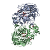
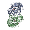
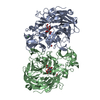
| Title | Two copies of the catalytic domain of NanA sialidase from Streptococcus pneumoniae juxtaposed in the P212121 space group, in complex with DANA derivatized with a PEG linker on the glycerol group. | ||||||
 Components Components | Sialidase A | ||||||
 Keywords Keywords | STRUCTURAL PROTEIN / Sialidase / Catalytic domain / CA170 | ||||||
| Function / homology |  Function and homology information Function and homology informationganglioside catabolic process / oligosaccharide catabolic process / exo-alpha-sialidase / exo-alpha-sialidase activity / membrane / cytoplasm Similarity search - Function | ||||||
| Biological species |   Streptococcus pneumoniae (bacteria) Streptococcus pneumoniae (bacteria) | ||||||
| Method |  X-RAY DIFFRACTION / SYNCHROTRON / MOLECULAR REPLACEMENT / Resolution: 1.94 Å X-RAY DIFFRACTION / SYNCHROTRON / MOLECULAR REPLACEMENT / Resolution: 1.94 Å | ||||||
 Authors Authors | Bridot, C. / Bouckaert, J. | ||||||
| Funding support |  France, 1items France, 1items
| ||||||
 Citation Citation | Journal: Chemistry / Year: 2021 Title: Polyvalent Transition-State Analogues of Sialyl Substrates Strongly Inhibit Bacterial Sialidases*. Authors: Assailly, C. / Bridot, C. / Saumonneau, A. / Lottin, P. / Roubinet, B. / Krammer, E.M. / Francois, F. / Vena, F. / Landemarre, L. / Alvarez Dorta, D. / Deniaud, D. / Grandjean, C. / Tellier, ...Authors: Assailly, C. / Bridot, C. / Saumonneau, A. / Lottin, P. / Roubinet, B. / Krammer, E.M. / Francois, F. / Vena, F. / Landemarre, L. / Alvarez Dorta, D. / Deniaud, D. / Grandjean, C. / Tellier, C. / Pascual, S. / Montembault, V. / Fontaine, L. / Daligault, F. / Bouckaert, J. / Gouin, S.G. #1: Journal: Chemistry / Year: 2019 Title: Multivalent Thiosialosides and Their Synergistic Interaction with Pathogenic Sialidases. Authors: Brissonnet, J. | ||||||
| History |
|
- Structure visualization
| Structure viewer | Molecule: MolmilJmol/JSmol |
|---|
- Downloads & links
-Download
| PDBx/mmCIF format | 7a5x.cif.gz | 382.5 KB | Display | PDBx/mmCIF format |
|---|---|---|---|---|
| PDB format | pdb7a5x.ent.gz | 307.3 KB | Display | PDB format |
| PDBx/mmJSON format | 7a5x.json.gz | Tree view | PDBx/mmJSON format | |
| Others |  Other downloads Other downloads |
-Validation report
| Arichive directory | https://data.pdbj.org/pub/pdb/validation_reports/a5/7a5xftp://data.pdbj.org/pub/pdb/validation_reports/a5/7a5x | HTTPS FTP |
|---|
-Related structure data
| Related structure data |  7a54C 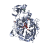 2ya5S S: Starting model for refinement C: citing same article ( |
|---|---|
| Similar structure data |
-Links
 PDBj
PDBj


- Assembly
| Deposited unit | 
| ||||||||||||
|---|---|---|---|---|---|---|---|---|---|---|---|---|---|
| 1 |
| ||||||||||||
| Unit cell |
|
-Components
-Protein , 1 types, 2 molecules AB
| #1: Protein | Mass: 56141.594 Da / Num. of mol.: 2 Source method: isolated from a genetically manipulated source Source: (gene. exp.) Streptococcus pneumoniae (bacteria) / Gene: nanA / Production host: |
|---|
-Non-polymers , 5 types, 756 molecules 


| #2: Chemical |  Mass: 462.518 Da / Num. of mol.: 2 / Source method: obtained synthetically / Formula: C18H30N4O8S Mass: 462.518 Da / Num. of mol.: 2 / Source method: obtained synthetically / Formula: C18H30N4O8S#3: Chemical |  Mass: 96.063 Da / Num. of mol.: 2 / Source method: obtained synthetically / Formula: SO4 Mass: 96.063 Da / Num. of mol.: 2 / Source method: obtained synthetically / Formula: SO4#4: Chemical | ChemComp-CL /  Mass: 35.453 Da / Num. of mol.: 4 / Source method: obtained synthetically / Formula: Cl Mass: 35.453 Da / Num. of mol.: 4 / Source method: obtained synthetically / Formula: Cl#5: Chemical |  Mass: 40.078 Da / Num. of mol.: 2 / Source method: obtained synthetically / Formula: Ca Mass: 40.078 Da / Num. of mol.: 2 / Source method: obtained synthetically / Formula: Ca#6: Water | ChemComp-HOH / | Mass: 18.015 Da / Num. of mol.: 746 / Source method: isolated from a natural source / Formula: H2O |
|---|
-Details
| Has ligand of interest | Y |
|---|
-Experimental details
-Experiment
| Experiment | Method: X-RAY DIFFRACTION / Number of used crystals: 1 |
|---|
- Sample preparation
| Crystal | Density Matthews: 2.4 Å3/Da / Density % sol: 48.73 % |
|---|---|
| Crystal grow | Temperature: 291 K / Method: vapor diffusion, sitting drop / pH: 8.5 Details: 0.2M Ammonium sulfate, 0.1M Tris pH=8,5 35% w/v PEG 3350 |
-Data collection
| Diffraction | Mean temperature: 100 K / Serial crystal experiment: N |
|---|---|
| Diffraction source | Source: SYNCHROTRON / Site: SOLEIL / Beamline: PROXIMA 1 / Wavelength: 0.987 Å |
| Detector | Type: DECTRIS EIGER X 16M / Detector: PIXEL / Date: Jan 25, 2019 |
| Radiation | Protocol: SINGLE WAVELENGTH / Monochromatic (M) / Laue (L): M / Scattering type: x-ray |
| Radiation wavelength | Wavelength: 0.987 Å / Relative weight: 1 |
| Reflection | Resolution: 1.94→56.6 Å / Num. obs: 80186 / % possible obs: 99.3 % / Redundancy: 8.43 % / Biso Wilson estimate: 38.564 Å2 / CC1/2: 0.997 / Rmerge(I) obs: 0.161 / Rrim(I) all: 0.171 / Net I/σ(I): 10.02 |
| Reflection shell | Resolution: 1.94→2.06 Å / Redundancy: 8.6 % / Rmerge(I) obs: 0.1628 / Mean I/σ(I) obs: 1.11 / Num. unique obs: 12318 / CC1/2: 0.477 / Rrim(I) all: 0.1731 / % possible all: 95.7 |
- Processing
| Software |
| ||||||||||||||||||||||||||||||||||||||||||||||||||||||||||||||||||||||||||||||||||||||||||||||||||||||||||||||||||||||||||||||||||||||||||||||||||||||||||||||||||||||||||||||||||||||||||||||||||||||||||||||||||
|---|---|---|---|---|---|---|---|---|---|---|---|---|---|---|---|---|---|---|---|---|---|---|---|---|---|---|---|---|---|---|---|---|---|---|---|---|---|---|---|---|---|---|---|---|---|---|---|---|---|---|---|---|---|---|---|---|---|---|---|---|---|---|---|---|---|---|---|---|---|---|---|---|---|---|---|---|---|---|---|---|---|---|---|---|---|---|---|---|---|---|---|---|---|---|---|---|---|---|---|---|---|---|---|---|---|---|---|---|---|---|---|---|---|---|---|---|---|---|---|---|---|---|---|---|---|---|---|---|---|---|---|---|---|---|---|---|---|---|---|---|---|---|---|---|---|---|---|---|---|---|---|---|---|---|---|---|---|---|---|---|---|---|---|---|---|---|---|---|---|---|---|---|---|---|---|---|---|---|---|---|---|---|---|---|---|---|---|---|---|---|---|---|---|---|---|---|---|---|---|---|---|---|---|---|---|---|---|---|---|---|---|
| Refinement | Method to determine structure: MOLECULAR REPLACEMENT Starting model: 2YA5 Resolution: 1.94→56.6 Å / SU ML: 0.2973 / Cross valid method: FREE R-VALUE / σ(F): 1.33 / Phase error: 26.0805 Stereochemistry target values: GeoStd + Monomer Library + CDL v1.2
| ||||||||||||||||||||||||||||||||||||||||||||||||||||||||||||||||||||||||||||||||||||||||||||||||||||||||||||||||||||||||||||||||||||||||||||||||||||||||||||||||||||||||||||||||||||||||||||||||||||||||||||||||||
| Solvent computation | Shrinkage radii: 0.9 Å / VDW probe radii: 1.11 Å / Solvent model: FLAT BULK SOLVENT MODEL / Bsol: 46.6 Å2 / ksol: 0.35 e/Å3 | ||||||||||||||||||||||||||||||||||||||||||||||||||||||||||||||||||||||||||||||||||||||||||||||||||||||||||||||||||||||||||||||||||||||||||||||||||||||||||||||||||||||||||||||||||||||||||||||||||||||||||||||||||
| Displacement parameters | Biso mean: 45.38 Å2 | ||||||||||||||||||||||||||||||||||||||||||||||||||||||||||||||||||||||||||||||||||||||||||||||||||||||||||||||||||||||||||||||||||||||||||||||||||||||||||||||||||||||||||||||||||||||||||||||||||||||||||||||||||
| Refinement step | Cycle: LAST / Resolution: 1.94→56.6 Å
| ||||||||||||||||||||||||||||||||||||||||||||||||||||||||||||||||||||||||||||||||||||||||||||||||||||||||||||||||||||||||||||||||||||||||||||||||||||||||||||||||||||||||||||||||||||||||||||||||||||||||||||||||||
| Refine LS restraints |
| ||||||||||||||||||||||||||||||||||||||||||||||||||||||||||||||||||||||||||||||||||||||||||||||||||||||||||||||||||||||||||||||||||||||||||||||||||||||||||||||||||||||||||||||||||||||||||||||||||||||||||||||||||
| LS refinement shell |
|
