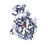
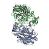
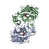
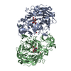
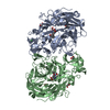
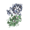
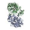
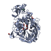
Mass: 55346.684 Da / Num. of mol.: 2 / Fragment: CATALYTIC DOMAIN, RESIDUES 280-754 Source method: isolated from a genetically manipulated source Details: THE SEQUENCE NUMBERING ABOVE CORRESPONDS TO THE CLOSEST UNIPROT SEQUENCE MATCH UNP B2DJD9. THE SEQUENCE NUMBERING FOR THE ENTRY SHOULD BE 303-777 Source: (gene. exp.) STREPTOCOCCUS PNEUMONIAE (bacteria) / Strain: TIGR4 / Production host: ESCHERICHIA COLI (E. coli) / Strain (production host): BL21 References: UniProt: B2DJD9, UniProt: P62575*PLUS, exo-alpha-sialidase
Mass: 18.015 Da / Num. of mol.: 838 / Source method: isolated from a natural source / Formula: H2O
Sequence details
HYPOTHETICAL PROTEIN SPNET_02001817 STREPTOCOCCUS PNEUMONIAE TIGR4. APPARENT CONFLICTS ARE DUE TO ...HYPOTHETICAL PROTEIN SPNET_02001817 STREPTOCOCCUS PNEUMONIAE TIGR4. APPARENT CONFLICTS ARE DUE TO CURRENT UNIPROT MAPPING. SEQUENCE REFERENCE: UNIREF100_UPI00005582E2.
-
Experimental details
-
Experiment
Experiment
Method: X-RAY DIFFRACTION / Number of used crystals: 1
-
Sample preparation
Crystal
Density Matthews: 2.22 Å3/Da / Density % sol: 44.5 % / Description: NONE
Crystal grow
Details: 20% PEG 3350, 200 MM KFORMATE, 20 MM TRIS PH 7.5 .
 Movie
Movie Controller


 Open data
 Basic information
 Components
 Keywords
 Function and homology information
 STREPTOCOCCUS PNEUMONIAE (bacteria)
 X-RAY DIFFRACTION /
  Authors
 Citation
 Structure visualization
 Downloads & links
 Other downloads
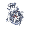


 PDBj
PDBj


 Assembly
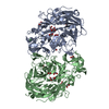

 Mass: 46.025 Da / Num. of mol.: 10 / Source method: obtained synthetically / Formula: CH2O2
 Mass: 62.068 Da / Num. of mol.: 6 / Source method: obtained synthetically / Formula: C2H6O2
 Mass: 35.453 Da / Num. of mol.: 2 / Source method: obtained synthetically / Formula: Cl
 Mass: 18.015 Da / Num. of mol.: 838 / Source method: isolated from a natural source / Formula: H2O
 Sample preparation
 / Beamline: BM14 / Wavelength: 0.976
  Processing