 Movie
Movie Controller

[English] 日本語
 Yorodumi
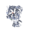
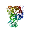
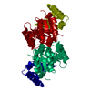
- PDB-2bf6: Atomic Resolution Structure of the bacterial sialidase NanI from ... -
+ Open data

- Basic information
| Entry | Database: PDB / ID: 2bf6 | ||||||
|---|---|---|---|---|---|---|---|
| Title | Atomic Resolution Structure of the bacterial sialidase NanI from Clostridium perfringens in complex with alpha-Sialic Acid (Neu5Ac). | ||||||
 Components Components | EXO-ALPHA-SIALIDASE | ||||||
 Keywords Keywords | HYDROLASE / SIALIDASE / CLOSTRIDIUM PERFRINGENS / SIALIC ACID | ||||||
| Function / homology |  Function and homology information Function and homology informationganglioside catabolic process / oligosaccharide catabolic process / exo-alpha-sialidase / exo-alpha-sialidase activity / membrane / metal ion binding / cytoplasm Similarity search - Function | ||||||
| Biological species |   Clostridium perfringens (bacteria) Clostridium perfringens (bacteria) | ||||||
| Method |  X-RAY DIFFRACTION / SYNCHROTRON / MOLECULAR REPLACEMENT / Resolution: 0.97 Å X-RAY DIFFRACTION / SYNCHROTRON / MOLECULAR REPLACEMENT / Resolution: 0.97 Å | ||||||
 Authors Authors | Newstead, S. / Taylor, G.L. | ||||||
 Citation Citation | Journal: J.Biol.Chem. / Year: 2008 Title: The Structure of Clostridium Perfringens Nani Sialidase and its Catalytic Intermediates. Authors: Newstead, S.L. / Potter, J.A. / Wilson, J.C. / Xu, G. / Chien, C.H. / Watts, A.G. / Withers, S.G. / Taylor, G.L. | ||||||
| History |
| ||||||
| Remark 700 | SHEET THE SHEET STRUCTURE OF THIS MOLECULE IS BIFURCATED. IN ORDER TO REPRESENT THIS FEATURE IN ... SHEET THE SHEET STRUCTURE OF THIS MOLECULE IS BIFURCATED. IN ORDER TO REPRESENT THIS FEATURE IN THE SHEET RECORDS BELOW, TWO SHEETS ARE DEFINED. |
- Structure visualization
| Structure viewer | Molecule: MolmilJmol/JSmol |
|---|
- Downloads & links
-Download
| PDBx/mmCIF format | 2bf6.cif.gz | 209 KB | Display | PDBx/mmCIF format |
|---|---|---|---|---|
| PDB format | pdb2bf6.ent.gz | 162 KB | Display | PDB format |
| PDBx/mmJSON format | 2bf6.json.gz | Tree view | PDBx/mmJSON format | |
| Others |  Other downloads Other downloads |
-Validation report
| Arichive directory | https://data.pdbj.org/pub/pdb/validation_reports/bf/2bf6ftp://data.pdbj.org/pub/pdb/validation_reports/bf/2bf6 | HTTPS FTP |
|---|
-Related structure data
| Related structure data | 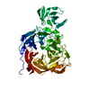 2vk5C  2vk6C 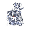 2vk7C 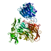 1sllS S: Starting model for refinement C: citing same article ( |
|---|---|
| Similar structure data |

-Links
 PDBj
PDBj

- Assembly
| Deposited unit | 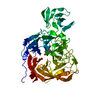
| ||||||||
|---|---|---|---|---|---|---|---|---|---|
| 1 |
| ||||||||
| Unit cell |
|
-Components
| #1: Protein | Mass: 50091.492 Da / Num. of mol.: 1 / Fragment: RESIDUES 243-691 Source method: isolated from a genetically manipulated source Source: (gene. exp.) Clostridium perfringens (bacteria) / Production host: | ||||
|---|---|---|---|---|---|
| #2: Sugar | ChemComp-SIA /   Type: D-saccharide, alpha linking / Mass: 309.270 Da / Num. of mol.: 1 Type: D-saccharide, alpha linking / Mass: 309.270 Da / Num. of mol.: 1Source method: isolated from a genetically manipulated source Formula: C11H19NO9 | ||||
| #3: Chemical |   Mass: 40.078 Da / Num. of mol.: 2 / Source method: obtained synthetically / Formula: Ca Mass: 40.078 Da / Num. of mol.: 2 / Source method: obtained synthetically / Formula: Ca#4: Chemical |   Mass: 92.094 Da / Num. of mol.: 2 / Source method: obtained synthetically / Formula: C3H8O3 Mass: 92.094 Da / Num. of mol.: 2 / Source method: obtained synthetically / Formula: C3H8O3#5: Water | ChemComp-HOH / |  Mass: 18.015 Da / Num. of mol.: 485 / Source method: isolated from a natural source / Formula: H2O Mass: 18.015 Da / Num. of mol.: 485 / Source method: isolated from a natural source / Formula: H2O |
-Experimental details
-Experiment
| Experiment | Method: X-RAY DIFFRACTION / Number of used crystals: 1 |
|---|
- Sample preparation
| Crystal | Density Matthews: 2.5 Å3/Da / Density % sol: 49 % |
|---|---|
| Crystal grow | pH: 7 / Details: 20 % PEG 3350, 0.2M POTASSIUM NITRATE, pH 7.00 |
-Data collection
| Diffraction | Mean temperature: 100 K |
|---|---|
| Diffraction source | Source: SYNCHROTRON / Site: ESRF  / Beamline: ID14-1 / Wavelength: 0.934 / Beamline: ID14-1 / Wavelength: 0.934 |
| Detector | Type: ADSC CCD / Detector: CCD / Date: Feb 20, 2004 / Details: MIRRORS |
| Radiation | Monochromator: DIAMOND (111), GE(220) / Protocol: SINGLE WAVELENGTH / Monochromatic (M) / Laue (L): M / Scattering type: x-ray |
| Radiation wavelength | Wavelength: 0.934 Å / Relative weight: 1 |
| Reflection | Resolution: 0.97→57 Å / Num. obs: 290992 / % possible obs: 91 % / Redundancy: 3.8 % / Rmerge(I) obs: 0.1 / Net I/σ(I): 11.8 |
| Reflection shell | Resolution: 0.97→1.03 Å / Redundancy: 3.1 % / Rmerge(I) obs: 0.19 / Mean I/σ(I) obs: 5 / % possible all: 87 |
- Processing
| Software |
| |||||||||||||||||||||||||||||||||
|---|---|---|---|---|---|---|---|---|---|---|---|---|---|---|---|---|---|---|---|---|---|---|---|---|---|---|---|---|---|---|---|---|---|---|
| Refinement | Method to determine structure: MOLECULAR REPLACEMENT Starting model: PDB ENTRY 1SLL Resolution: 0.97→57 Å / Num. parameters: 36804 / Num. restraintsaints: 44962 / Cross valid method: FREE R-VALUE / σ(F): 0 / Stereochemistry target values: ENGH AND HUBER Details: ANISOTROPIC REFINEMENT REDUCED FREE R (NO CUTOFF) BY 2.0
| |||||||||||||||||||||||||||||||||
| Refine analyze | Num. disordered residues: 11 / Occupancy sum hydrogen: 3285.2 / Occupancy sum non hydrogen: 4037.5 | |||||||||||||||||||||||||||||||||
| Refinement step | Cycle: LAST / Resolution: 0.97→57 Å
| |||||||||||||||||||||||||||||||||
| Refine LS restraints |
|
