 Movie
Movie Controller

[English] 日本語
 Yorodumi
- PDB-3g1h: Crystal structure of orotidine 5'-monophosphate decarboxylase fro... -
+ Open data

- Basic information
| Entry | Database: PDB / ID: 3g1h | ||||||
|---|---|---|---|---|---|---|---|
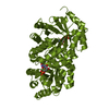
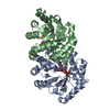
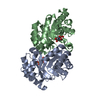
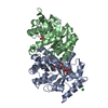
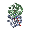
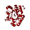
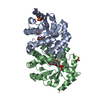
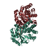
| Title | Crystal structure of orotidine 5'-monophosphate decarboxylase from Methanobacterium thermoautotrophicum complexed with 5,6-dihydrouridine 5'-monophosphate | ||||||
 Components Components | Orotidine 5'-phosphate decarboxylase | ||||||
 Keywords Keywords | LYASE / orotidine 5'-monophosphate decarboxylase / H2-UMP / Decarboxylase / Pyrimidine biosynthesis | ||||||
| Function / homology |  Function and homology information Function and homology informationorotidine-5'-phosphate decarboxylase / orotidine-5'-phosphate decarboxylase activity / 'de novo' UMP biosynthetic process / 'de novo' pyrimidine nucleobase biosynthetic process / cytosol Similarity search - Function | ||||||
| Biological species |   Methanothermobacter thermautotrophicus str. Delta H (archaea) Methanothermobacter thermautotrophicus str. Delta H (archaea) | ||||||
| Method |  X-RAY DIFFRACTION / SYNCHROTRON / MOLECULAR REPLACEMENT / Resolution: 2.3 Å X-RAY DIFFRACTION / SYNCHROTRON / MOLECULAR REPLACEMENT / Resolution: 2.3 Å | ||||||
 Authors Authors | Fedorov, A.A. / Fedorov, E.V. / Chan, K.K. / Gerlt, J.A. / Almo, S.C. | ||||||
 Citation Citation | Journal: Biochemistry / Year: 2009 Title: Mechanism of the orotidine 5'-monophosphate decarboxylase-catalyzed reaction: evidence for substrate destabilization. Authors: Chan, K.K. / Wood, B.M. / Fedorov, A.A. / Fedorov, E.V. / Imker, H.J. / Amyes, T.L. / Richard, J.P. / Almo, S.C. / Gerlt, J.A. | ||||||
| History |
|
- Structure visualization
| Structure viewer | Molecule: MolmilJmol/JSmol |
|---|
- Downloads & links
-Download
| PDBx/mmCIF format | 3g1h.cif.gz | 533.5 KB | Display | PDBx/mmCIF format |
|---|---|---|---|---|
| PDB format | pdb3g1h.ent.gz | 441.1 KB | Display | PDB format |
| PDBx/mmJSON format | 3g1h.json.gz | Tree view | PDBx/mmJSON format | |
| Others |  Other downloads Other downloads |
-Validation report
| Arichive directory | https://data.pdbj.org/pub/pdb/validation_reports/g1/3g1hftp://data.pdbj.org/pub/pdb/validation_reports/g1/3g1h | HTTPS FTP |
|---|
-Related structure data
| Related structure data |  3g18SC  3g1aC  3g1dC 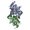 3g1fC  3g1sC 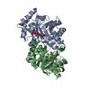 3g1vC  3g1xC  3g1yC  3g22C  3g24C  3gdkC  3gdlC  3gdrC  3gdtC S: Starting model for refinement C: citing same article ( |
|---|---|
| Similar structure data |


-Links
 PDBj
PDBj
- Assembly
| Deposited unit | 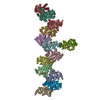
| ||||||||
|---|---|---|---|---|---|---|---|---|---|
| 1 | 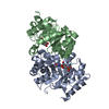
| ||||||||
| 2 | 
| ||||||||
| 3 | 
| ||||||||
| 4 | 
| ||||||||
| 5 | 
| ||||||||
| 6 | 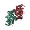
| ||||||||
| 7 | 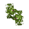
| ||||||||
| Unit cell |
| ||||||||
| Components on special symmetry positions |
|
-Components
| #1: Protein | Mass: 24884.697 Da / Num. of mol.: 13 / Mutation: R101P Source method: isolated from a genetically manipulated source Source: (gene. exp.) Methanothermobacter thermautotrophicus str. Delta H (archaea)Gene: pyrF, MTH_129 / Production host:  References: UniProt: O26232, orotidine-5'-phosphate decarboxylase #2: Chemical | ChemComp-H2U /   Type: RNA linking / Mass: 326.197 Da / Num. of mol.: 13 / Source method: obtained synthetically / Formula: C9H15N2O9P Type: RNA linking / Mass: 326.197 Da / Num. of mol.: 13 / Source method: obtained synthetically / Formula: C9H15N2O9P#3: Water | ChemComp-HOH / |  Mass: 18.015 Da / Num. of mol.: 246 / Source method: isolated from a natural source / Formula: H2O Mass: 18.015 Da / Num. of mol.: 246 / Source method: isolated from a natural source / Formula: H2OHas protein modification | Y | |
|---|
-Experimental details
-Experiment
| Experiment | Method: X-RAY DIFFRACTION / Number of used crystals: 1 |
|---|
- Sample preparation
| Crystal | Density Matthews: 2.24 Å3/Da / Density % sol: 45.16 % |
|---|---|
| Crystal grow | Temperature: 293 K / Method: vapor diffusion, hanging drop / pH: 5.6 Details: 30% PEG 4000, 0.1M sodium citrate, 0.2M ammonium acetate, pH 5.6, VAPOR DIFFUSION, HANGING DROP, temperature 293K |
-Data collection
| Diffraction | Mean temperature: 100 K |
|---|---|
| Diffraction source | Source: SYNCHROTRON / Site: NSLS  / Beamline: X4A / Wavelength: 0.97915 Å / Beamline: X4A / Wavelength: 0.97915 Å |
| Detector | Type: ADSC QUANTUM 4 / Detector: CCD / Date: Mar 21, 2008 |
| Radiation | Monochromator: Si 111 CHANNEL / Protocol: SINGLE WAVELENGTH / Monochromatic (M) / Laue (L): M / Scattering type: x-ray |
| Radiation wavelength | Wavelength: 0.97915 Å / Relative weight: 1 |
| Reflection | Resolution: 2.3→25 Å / Num. all: 126808 / Num. obs: 126808 / % possible obs: 100 % / Observed criterion σ(F): 0 / Observed criterion σ(I): 0 / Biso Wilson estimate: 41.8 Å2 / Rmerge(I) obs: 0.067 |
- Processing
| Software |
| ||||||||||||||||||||||||||||||||||||
|---|---|---|---|---|---|---|---|---|---|---|---|---|---|---|---|---|---|---|---|---|---|---|---|---|---|---|---|---|---|---|---|---|---|---|---|---|---|
| Refinement | Method to determine structure: MOLECULAR REPLACEMENT Starting model: 3G18 Resolution: 2.3→24.76 Å / Rfactor Rfree error: 0.004 / Data cutoff high absF: 1916415.64 / Data cutoff low absF: 0 / Isotropic thermal model: RESTRAINED / Cross valid method: THROUGHOUT / σ(F): 0 / σ(I): 0 / Stereochemistry target values: Engh & Huber
| ||||||||||||||||||||||||||||||||||||
| Solvent computation | Solvent model: FLAT MODEL / Bsol: 38.8084 Å2 / ksol: 0.331807 e/Å3 | ||||||||||||||||||||||||||||||||||||
| Displacement parameters | Biso mean: 64 Å2
| ||||||||||||||||||||||||||||||||||||
| Refine analyze |
| ||||||||||||||||||||||||||||||||||||
| Refinement step | Cycle: LAST / Resolution: 2.3→24.76 Å
| ||||||||||||||||||||||||||||||||||||
| Refine LS restraints |
| ||||||||||||||||||||||||||||||||||||
| LS refinement shell | Resolution: 2.3→2.38 Å / Rfactor Rfree error: 0.015 / Total num. of bins used: 10
| ||||||||||||||||||||||||||||||||||||
| Xplor file |
|
