regulation of translation / large ribosomal subunit / tRNA binding / rRNA binding / structural constituent of ribosome / translation Similarity search - Function
50S ribosomal protein L1; Chain A, Domain 1 / Arc Repressor Mutant, subunit A / Ribosomal protein L1, bacterial-type / Ribosomal protein L1, conserved site / Ribosomal protein L1 signature. / Ribosomal protein L1 / Ribosomal protein L1, 3-layer alpha/beta-sandwich / Ribosomal protein L1-like / Ribosomal protein L1/ribosomal biogenesis protein / Ribosomal protein L1p/L10e family ...50S ribosomal protein L1; Chain A, Domain 1 / Arc Repressor Mutant, subunit A / Ribosomal protein L1, bacterial-type / Ribosomal protein L1, conserved site / Ribosomal protein L1 signature. / Ribosomal protein L1 / Ribosomal protein L1, 3-layer alpha/beta-sandwich / Ribosomal protein L1-like / Ribosomal protein L1/ribosomal biogenesis protein / Ribosomal protein L1p/L10e family / Helix non-globular / Special Similarity search - Domain/homology
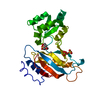
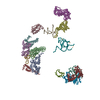
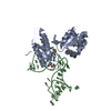
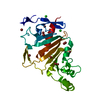
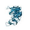
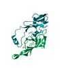
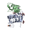
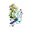
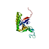
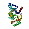
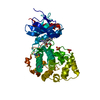
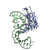
A: 50S RIBOSOMAL PROTEIN L1 B: FRAGMENT OF MRNA FOR L1-OPERON CONTAINING REGULATOR L1-BINDING SITE C: 50S RIBOSOMAL PROTEIN L1 D: FRAGMENT OF MRNA FOR L1-OPERON CONTAINING REGULATOR L1-BINDING SITE hetero molecules
Mass: 15522.342 Da / Num. of mol.: 2 Source method: isolated from a genetically manipulated source Source: (gene. exp.) METHANOCALDOCOCCUS JANNASCHII (archaea) Production host: ESCHERICHIA COLI (E. coli)
 Movie
Movie Controller

 Yorodumi
 Open data
 Basic information
 Components
 Keywords
 Function and homology information
 THERMUS THERMOPHILUS (bacteria)
 X-RAY DIFFRACTION /
  Authors
 Citation
 Structure visualization
 Downloads & links
 Other downloads
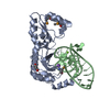


 PDBj
PDBj


 Assembly


 Mass: 39.098 Da / Num. of mol.: 2 / Source method: obtained synthetically / Formula: K
 Mass: 18.015 Da / Num. of mol.: 318 / Source method: isolated from a natural source / Formula: H2O
 Sample preparation
 / Beamline: BW7B / Wavelength: 0.8423
  Processing