homoserine O-succinyltransferase activity / L-methionine biosynthetic process from homoserine via O-succinyl-L-homoserine and cystathionine / homoserine O-acetyltransferase / homoserine O-acetyltransferase activity / cytoplasm Similarity search - Function
Homoserine O-succinyltransferase MetA / MetA family / Homoserine O-succinyltransferase / Class I glutamine amidotransferase (GATase) domain / Class I glutamine amidotransferase-like / Rossmann fold / 3-Layer(aba) Sandwich / Alpha Beta Similarity search - Domain/homology
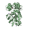
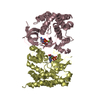
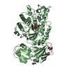
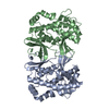
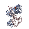
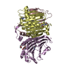
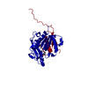
Mass: 35392.133 Da / Num. of mol.: 1 Source method: isolated from a genetically manipulated source Details: PROTEIN IS A PHYSIOLOGICAL DIMER, DIMER FORMED BY CRYSTALLOGRAPHIC TWO-FOLD Source: (gene. exp.) BACILLUS CEREUS (bacteria) / Production host: ESCHERICHIA COLI (E. coli) / Variant (production host): BL-21 CODON PLUS References: UniProt: Q72X44, homoserine O-succinyltransferase
Mass: 18.015 Da / Num. of mol.: 163 / Source method: isolated from a natural source / Formula: H2O
Nonpolymer details
SULFATE ION (SO4): SULFATE FROM CRYSTALLIZATION CONDITIONS L-HOMOSERINE (HSE): CO-CRYSTALLIZED WITH ...SULFATE ION (SO4): SULFATE FROM CRYSTALLIZATION CONDITIONS L-HOMOSERINE (HSE): CO-CRYSTALLIZED WITH HOMOSERINE 10MM
Sequence details
RESIDUES 1-16 AND 298-301 WERE NOT VISIBLE IN XTAL STRUCTURE
-
Experimental details
-
Experiment
Experiment
Method: X-RAY DIFFRACTION / Number of used crystals: 1
-
Sample preparation
Crystal
Density Matthews: 2.5 Å3/Da / Density % sol: 49 % / Description: NONE
 Movie
Movie Controller

 Yorodumi
 Open data
 Basic information
 Components
 Keywords
 Function and homology information
 X-RAY DIFFRACTION /
  Authors
 Citation
 Structure visualization
 Downloads & links
 Other downloads
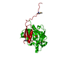


 PDBj
PDBj

 Assembly
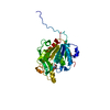

 Mass: 96.063 Da / Num. of mol.: 1 / Source method: obtained synthetically / Formula: SO4
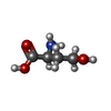
 Type: L-peptide linking / Mass: 119.119 Da / Num. of mol.: 1 / Source method: obtained synthetically / Formula: C4H9NO3
 Mass: 18.015 Da / Num. of mol.: 163 / Source method: isolated from a natural source / Formula: H2O
 Sample preparation
 / Beamline: BL9-1 / Wavelength: 0.979
  Processing