 Movie
Movie Controller

[English] 日本語
 Yorodumi
- PDB-6bm7: Crystal structure of Trypanosoma brucei AdoMetDC/prozyme heterodi... -
+ Open data

- Basic information
| Entry | Database: PDB / ID: 6bm7 | |||||||||
|---|---|---|---|---|---|---|---|---|---|---|
| Title | Crystal structure of Trypanosoma brucei AdoMetDC/prozyme heterodimer in complex with pyrimidineamine inhibitor UTSAM568 | |||||||||
 Components Components |
| |||||||||
 Keywords Keywords | LYASE/LYASE INHIBITOR / AdoMetDC / prozyme / decarboxylase / Trypanosoma / inhibitor / LYASE-LYASE INHIBITOR complex | |||||||||
| Function / homology |  Function and homology information Function and homology informationtrypanothione biosynthetic process / positive regulation of spermidine biosynthetic process / spermine biosynthetic process / adenosylmethionine decarboxylase / adenosylmethionine decarboxylase activity / positive regulation of catalytic activity / spermidine biosynthetic process / S-adenosylmethionine metabolic process / catalytic complex / enzyme activator activity ...trypanothione biosynthetic process / positive regulation of spermidine biosynthetic process / spermine biosynthetic process / adenosylmethionine decarboxylase / adenosylmethionine decarboxylase activity / positive regulation of catalytic activity / spermidine biosynthetic process / S-adenosylmethionine metabolic process / catalytic complex / enzyme activator activity / protein heterodimerization activity / enzyme binding / cytosol Similarity search - Function | |||||||||
| Biological species |  | |||||||||
| Method |  X-RAY DIFFRACTION / SYNCHROTRON / MOLECULAR REPLACEMENT / Resolution: 2.98 Å X-RAY DIFFRACTION / SYNCHROTRON / MOLECULAR REPLACEMENT / Resolution: 2.98 Å | |||||||||
 Authors Authors | Volkov, O.A. / Chen, Z. / Phillips, M.A. | |||||||||
| Funding support |  United States, 2items United States, 2items
| |||||||||
 Citation Citation | Journal: J. Med. Chem. / Year: 2018 Title: Species-Selective Pyrimidineamine Inhibitors of Trypanosoma brucei S-Adenosylmethionine Decarboxylase. Authors: Volkov, O.A. / Brockway, A.J. / Wring, S.A. / Peel, M. / Chen, Z. / Phillips, M.A. / De Brabander, J.K. | |||||||||
| History |
|
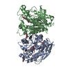
- Structure visualization
| Structure viewer | Molecule: MolmilJmol/JSmol |
|---|
- Downloads & links
-Download
| PDBx/mmCIF format | 6bm7.cif.gz | 593.3 KB | Display | PDBx/mmCIF format |
|---|---|---|---|---|
| PDB format | pdb6bm7.ent.gz | 395.8 KB | Display | PDB format |
| PDBx/mmJSON format | 6bm7.json.gz | Tree view | PDBx/mmJSON format | |
| Others |  Other downloads Other downloads |
-Validation report
| Arichive directory | https://data.pdbj.org/pub/pdb/validation_reports/bm/6bm7ftp://data.pdbj.org/pub/pdb/validation_reports/bm/6bm7 | HTTPS FTP |
|---|
-Related structure data
| Related structure data |  5tvmS S: Starting model for refinement |
|---|---|
| Similar structure data |


-Links
 PDBj
PDBj- Assembly
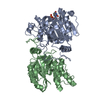
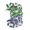
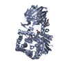
| Deposited unit | 
| ||||||||||||
|---|---|---|---|---|---|---|---|---|---|---|---|---|---|
| 1 | 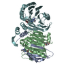
| ||||||||||||
| 2 | 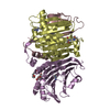
| ||||||||||||
| Unit cell |
|
-Components
-S-adenosylmethionine decarboxylase ... , 2 types, 4 molecules ACBD
| #1: Protein | Mass: 9754.132 Da / Num. of mol.: 2 / Fragment: UNP residues 1-85 Source method: isolated from a genetically manipulated source Source: (gene. exp.) Strain: 927/4 GUTat10.1 / Gene: Tb927.6.4410, Tb927.6.4460 / Plasmid: pETDuet-1 / Production host:  References: UniProt: Q587A7, adenosylmethionine decarboxylase #2: Protein | Mass: 32041.992 Da / Num. of mol.: 2 / Fragment: UNP residues 87-370 Source method: isolated from a genetically manipulated source Source: (gene. exp.) Strain: 927/4 GUTat10.1 / Gene: Tb927.6.4410, Tb927.6.4460 / Plasmid: pETDuet-1 / Production host: References: UniProt: Q587A7, adenosylmethionine decarboxylase |
|---|
-Protein , 1 types, 2 molecules EF
| #3: Protein | Mass: 36343.695 Da / Num. of mol.: 2 Source method: isolated from a genetically manipulated source Source: (gene. exp.) Strain: 927/4 GUTat10.1 / Plasmid: pETDuet-1 / Production host: |
|---|
-Non-polymers , 6 types, 16 molecules 

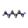


| #4: Chemical |  Mass: 282.334 Da / Num. of mol.: 2 / Source method: obtained synthetically / Formula: C11H26N2O6 / Comment: pH buffer*YM Mass: 282.334 Da / Num. of mol.: 2 / Source method: obtained synthetically / Formula: C11H26N2O6 / Comment: pH buffer*YM#5: Chemical |  Mass: 359.040 Da / Num. of mol.: 2 / Source method: obtained synthetically / Formula: C11H11Br2N4 / Feature type: SUBJECT OF INVESTIGATION Mass: 359.040 Da / Num. of mol.: 2 / Source method: obtained synthetically / Formula: C11H11Br2N4 / Feature type: SUBJECT OF INVESTIGATION#6: Chemical |  Mass: 88.151 Da / Num. of mol.: 2 / Source method: obtained synthetically / Formula: C4H12N2 Mass: 88.151 Da / Num. of mol.: 2 / Source method: obtained synthetically / Formula: C4H12N2#7: Chemical |  Mass: 150.173 Da / Num. of mol.: 2 / Source method: obtained synthetically / Formula: C6H14O4 Mass: 150.173 Da / Num. of mol.: 2 / Source method: obtained synthetically / Formula: C6H14O4#8: Chemical | ChemComp-PG4 / |  Mass: 194.226 Da / Num. of mol.: 1 / Source method: obtained synthetically / Formula: C8H18O5 / Comment: precipitant*YM Mass: 194.226 Da / Num. of mol.: 1 / Source method: obtained synthetically / Formula: C8H18O5 / Comment: precipitant*YM#9: Water | ChemComp-HOH / | Mass: 18.015 Da / Num. of mol.: 7 / Source method: isolated from a natural source / Formula: H2O |
|---|
-Details
| Has protein modification | Y |
|---|
-Experimental details
-Experiment
| Experiment | Method: X-RAY DIFFRACTION / Number of used crystals: 1 |
|---|
- Sample preparation
| Crystal | Density Matthews: 2.6 Å3/Da / Density % sol: 52.63 % / Description: plate |
|---|---|
| Crystal grow | Temperature: 293 K / Method: vapor diffusion, hanging drop / pH: 7 Details: 1 uL 4 mg/mL protein in crystallization buffer (50 mM Bis-Tris propane, pH 7.2, 50 mM sodium chloride, 4 mM TCEP, 2 mM putrescine, 0.5 mM N4-(3,5-dibromophenyl)-6-methylpyrimidine-2,4- ...Details: 1 uL 4 mg/mL protein in crystallization buffer (50 mM Bis-Tris propane, pH 7.2, 50 mM sodium chloride, 4 mM TCEP, 2 mM putrescine, 0.5 mM N4-(3,5-dibromophenyl)-6-methylpyrimidine-2,4-diamine [UTSAM568], 1% DMSO) + 1 uL reservoir solution (17% PEG6000, 100 mM Bis-Tris propane, pH 7.0) + 0.5 uL unliganded protein microseeds obtained using Seed-Bead method in stabilization solution (100 mM Bis-Tris propane, pH 8.9, 50 mM HEPES, pH 7.2, 19% PEG6000, 50 mM sodium chloride, 4 mM TCEP, 2 mM putrescine) |
-Data collection
| Diffraction | Mean temperature: 100 K |
|---|---|
| Diffraction source | Source: SYNCHROTRON / Site: APS / Beamline: 19-ID / Wavelength: 0.91905 Å |
| Detector | Type: ADSC QUANTUM 315r / Detector: CCD / Date: Dec 10, 2015 |
| Radiation | Monochromator: double crystal Si(111) / Protocol: SINGLE WAVELENGTH / Monochromatic (M) / Laue (L): M / Scattering type: x-ray |
| Radiation wavelength | Wavelength: 0.91905 Å / Relative weight: 1 |
| Reflection | Resolution: 2.98→50 Å / Num. obs: 30294 / % possible obs: 99.8 % / Redundancy: 6.8 % / Biso Wilson estimate: 49.2238648407 Å2 / Rpim(I) all: 0.074 / Rrim(I) all: 0.194 / Net I/σ(I): 10 |
| Reflection shell | Resolution: 3→3.05 Å / Redundancy: 6.3 % / Mean I/σ(I) obs: 1.87 / Num. unique obs: 1466 / CC1/2: 0.712 / % possible all: 99.1 |
- Processing
| Software |
| |||||||||||||||||||||||||||||||||||||||||||||||||||||||||||||||||||||||||||||||||||||||||||||||||||||||||||||||||||||||||||||||||||||
|---|---|---|---|---|---|---|---|---|---|---|---|---|---|---|---|---|---|---|---|---|---|---|---|---|---|---|---|---|---|---|---|---|---|---|---|---|---|---|---|---|---|---|---|---|---|---|---|---|---|---|---|---|---|---|---|---|---|---|---|---|---|---|---|---|---|---|---|---|---|---|---|---|---|---|---|---|---|---|---|---|---|---|---|---|---|---|---|---|---|---|---|---|---|---|---|---|---|---|---|---|---|---|---|---|---|---|---|---|---|---|---|---|---|---|---|---|---|---|---|---|---|---|---|---|---|---|---|---|---|---|---|---|---|---|
| Refinement | Method to determine structure: MOLECULAR REPLACEMENT Starting model: PDB entry 5TVM Resolution: 2.98→48.2623233532 Å / SU ML: 0.341675147008 / Cross valid method: FREE R-VALUE / σ(F): 1.33789805147 / Phase error: 26.3118648791
| |||||||||||||||||||||||||||||||||||||||||||||||||||||||||||||||||||||||||||||||||||||||||||||||||||||||||||||||||||||||||||||||||||||
| Solvent computation | Shrinkage radii: 0.9 Å / VDW probe radii: 1.11 Å | |||||||||||||||||||||||||||||||||||||||||||||||||||||||||||||||||||||||||||||||||||||||||||||||||||||||||||||||||||||||||||||||||||||
| Displacement parameters | Biso mean: 58.4151476361 Å2 | |||||||||||||||||||||||||||||||||||||||||||||||||||||||||||||||||||||||||||||||||||||||||||||||||||||||||||||||||||||||||||||||||||||
| Refinement step | Cycle: LAST / Resolution: 2.98→48.2623233532 Å
| |||||||||||||||||||||||||||||||||||||||||||||||||||||||||||||||||||||||||||||||||||||||||||||||||||||||||||||||||||||||||||||||||||||
| Refine LS restraints |
| |||||||||||||||||||||||||||||||||||||||||||||||||||||||||||||||||||||||||||||||||||||||||||||||||||||||||||||||||||||||||||||||||||||
| LS refinement shell |
|
