 Movie
Movie Controller

[English] 日本語
 Yorodumi
- PDB-5wt1: Pyrococcus abyssi methyltransferase PaTrm5a bound by SAH and cogn... -
+ Open data

- Basic information
| Entry | Database: PDB / ID: 5wt1 | ||||||
|---|---|---|---|---|---|---|---|
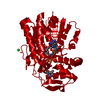
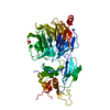
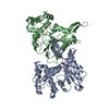
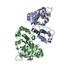
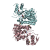
| Title | Pyrococcus abyssi methyltransferase PaTrm5a bound by SAH and cognate tRNA | ||||||
 Components Components |
| ||||||
 Keywords Keywords | TRANSFERASE/RNA / methyltransferase / Trm5a / wyosine hypermodification / TRANSFERASE-RNA complex | ||||||
| Function / homology |  Function and homology information Function and homology informationtRNA (guanine37-N1)-methyltransferase / tRNA (guanine(37)-N1)-methyltransferase activity / tRNA N1-guanine methylation / tRNA methylation / Transferases; Transferring one-carbon groups; Methyltransferases / cytoplasm Similarity search - Function | ||||||
| Biological species |   Pyrococcus abyssi (archaea)Pyrococcus abyssi GE5 (archaea) Pyrococcus abyssi (archaea)Pyrococcus abyssi GE5 (archaea) | ||||||
| Method |  X-RAY DIFFRACTION / SYNCHROTRON / MOLECULAR REPLACEMENT / Resolution: 2.598 Å X-RAY DIFFRACTION / SYNCHROTRON / MOLECULAR REPLACEMENT / Resolution: 2.598 Å | ||||||
 Authors Authors | Xie, W. / Wang, C. / Jia, Q. | ||||||
 Citation Citation | Journal: Sci Adv / Year: 2017 Title: Structural insight into the methyltransfer mechanism of the bifunctional Trm5. Authors: Wang, C. / Jia, Q. / Zeng, J. / Chen, R. / Xie, W. | ||||||
| History |
|
- Structure visualization
| Structure viewer | Molecule: MolmilJmol/JSmol |
|---|
- Downloads & links
-Download
| PDBx/mmCIF format | 5wt1.cif.gz | 215.9 KB | Display | PDBx/mmCIF format |
|---|---|---|---|---|
| PDB format | pdb5wt1.ent.gz | 164.8 KB | Display | PDB format |
| PDBx/mmJSON format | 5wt1.json.gz | Tree view | PDBx/mmJSON format | |
| Others |  Other downloads Other downloads |
-Validation report
| Arichive directory | https://data.pdbj.org/pub/pdb/validation_reports/wt/5wt1ftp://data.pdbj.org/pub/pdb/validation_reports/wt/5wt1 | HTTPS FTP |
|---|
-Related structure data
| Related structure data |  5wt3C  2zznS C: citing same article ( S: Starting model for refinement |
|---|---|
| Similar structure data |


-Links
 PDBj
PDBj


- Assembly
| Deposited unit | 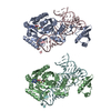
| ||||||||
|---|---|---|---|---|---|---|---|---|---|
| 1 | 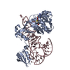
| ||||||||
| 2 | 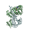
| ||||||||
| Unit cell |
|
-Components
| #1: Protein | Mass: 38545.062 Da / Num. of mol.: 2 Source method: isolated from a genetically manipulated source Source: (gene. exp.) Pyrococcus abyssi (strain GE5 / Orsay) (archaea)Strain: GE5 / Orsay / Gene: trm5a, PYRAB01130, PAB2272 / Production host:  References: UniProt: Q9V2G1, tRNA (guanine37-N1)-methyltransferase #2: RNA chain | Mass: 24551.658 Da / Num. of mol.: 2 / Source method: obtained synthetically / Source: (synth.) Pyrococcus abyssi GE5 (archaea) / References: GenBank: HE613800.1#3: Chemical |   Type: L-peptide linking / Mass: 384.411 Da / Num. of mol.: 2 / Source method: obtained synthetically / Formula: C14H20N6O5S Type: L-peptide linking / Mass: 384.411 Da / Num. of mol.: 2 / Source method: obtained synthetically / Formula: C14H20N6O5S#4: Water | ChemComp-HOH / |  Mass: 18.015 Da / Num. of mol.: 60 / Source method: isolated from a natural source / Formula: H2O Mass: 18.015 Da / Num. of mol.: 60 / Source method: isolated from a natural source / Formula: H2O |
|---|
-Experimental details
-Experiment
| Experiment | Method: X-RAY DIFFRACTION / Number of used crystals: 1 |
|---|
- Sample preparation
| Crystal | Density Matthews: 2.62 Å3/Da / Density % sol: 53 % |
|---|---|
| Crystal grow | Temperature: 298 K / Method: vapor diffusion, sitting drop / pH: 6.5 / Details: 45% MPD, 100 mM MES (pH 6.5), 200 mM NH4OAc |
-Data collection
| Diffraction | Mean temperature: 100 K |
|---|---|
| Diffraction source | Source: SYNCHROTRON / Site: SSRF  / Beamline: BL17U / Wavelength: 0.99 Å / Beamline: BL17U / Wavelength: 0.99 Å |
| Detector | Type: ADSC QUANTUM 315r / Detector: CCD / Date: Apr 2, 2016 |
| Radiation | Protocol: SINGLE WAVELENGTH / Monochromatic (M) / Laue (L): M / Scattering type: x-ray |
| Radiation wavelength | Wavelength: 0.99 Å / Relative weight: 1 |
| Reflection | Resolution: 2.598→50 Å / Num. obs: 40587 / % possible obs: 99.5 % / Redundancy: 3.7 % / Rmerge(I) obs: 0.077 / Net I/σ(I): 15.6 |
| Reflection shell | Resolution: 2.6→2.69 Å / Redundancy: 3.8 % / Rmerge(I) obs: 0.911 / Mean I/σ(I) obs: 1.7 / CC1/2: 0.739 / % possible all: 99.9 |
- Processing
| Software |
| |||||||||||||||||||||||||||||||||||||||||||||||||||||||||||||||||||||||||||||||||||||||||||||||||||||||||
|---|---|---|---|---|---|---|---|---|---|---|---|---|---|---|---|---|---|---|---|---|---|---|---|---|---|---|---|---|---|---|---|---|---|---|---|---|---|---|---|---|---|---|---|---|---|---|---|---|---|---|---|---|---|---|---|---|---|---|---|---|---|---|---|---|---|---|---|---|---|---|---|---|---|---|---|---|---|---|---|---|---|---|---|---|---|---|---|---|---|---|---|---|---|---|---|---|---|---|---|---|---|---|---|---|---|---|
| Refinement | Method to determine structure: MOLECULAR REPLACEMENT Starting model: 2ZZN Resolution: 2.598→40.217 Å / SU ML: 0.47 / Cross valid method: FREE R-VALUE / σ(F): 1.34 / Phase error: 34.31 / Stereochemistry target values: ML
| |||||||||||||||||||||||||||||||||||||||||||||||||||||||||||||||||||||||||||||||||||||||||||||||||||||||||
| Solvent computation | Shrinkage radii: 0.9 Å / VDW probe radii: 1.11 Å / Solvent model: FLAT BULK SOLVENT MODEL | |||||||||||||||||||||||||||||||||||||||||||||||||||||||||||||||||||||||||||||||||||||||||||||||||||||||||
| Refinement step | Cycle: LAST / Resolution: 2.598→40.217 Å
| |||||||||||||||||||||||||||||||||||||||||||||||||||||||||||||||||||||||||||||||||||||||||||||||||||||||||
| Refine LS restraints |
| |||||||||||||||||||||||||||||||||||||||||||||||||||||||||||||||||||||||||||||||||||||||||||||||||||||||||
| LS refinement shell |
|
