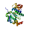
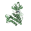
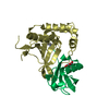
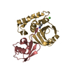
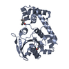
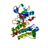
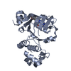
TRANSPORT PROTEIN / ABCC6 / NBD2 / ADP-bound state / ATP-Binding Cassette transporter C6 / nucleotide binding domain 2
Function / homology
Function and homology information
Defective ABCC6 causes PXE / inorganic diphosphate transport / inhibition of non-skeletal tissue mineralization / ATP transport / response to sodium phosphate / leukotriene transport / ABC-type glutathione-S-conjugate transporter / phosphate ion homeostasis / ABC-type glutathione S-conjugate transporter activity / : ...Defective ABCC6 causes PXE / inorganic diphosphate transport / inhibition of non-skeletal tissue mineralization / ATP transport / response to sodium phosphate / leukotriene transport / ABC-type glutathione-S-conjugate transporter / phosphate ion homeostasis / ABC-type glutathione S-conjugate transporter activity / : / intracellular phosphate ion homeostasis / Translocases; Catalysing the translocation of other compounds; Linked to the hydrolysis of a nucleoside triphosphate / ATPase-coupled transmembrane transporter activity / response to magnesium ion / ABC-type transporter activity / ATP metabolic process / calcium ion homeostasis / visual perception / basal plasma membrane / transmembrane transport / ABC-family protein mediated transport / gene expression / basolateral plasma membrane / response to xenobiotic stimulus / endoplasmic reticulum membrane / ATP hydrolysis activity / extracellular region / nucleoplasm / ATP binding / plasma membrane Similarity search - Function
Multi drug resistance-associated protein / : / ABC transporter TMD0 domain / : / ABC transporter transmembrane region / ABC transporter type 1, transmembrane domain / ABC transporter integral membrane type-1 fused domain profile. / ABC transporter type 1, transmembrane domain superfamily / ABC transporter-like, conserved site / ABC transporters family signature. ...Multi drug resistance-associated protein / : / ABC transporter TMD0 domain / : / ABC transporter transmembrane region / ABC transporter type 1, transmembrane domain / ABC transporter integral membrane type-1 fused domain profile. / ABC transporter type 1, transmembrane domain superfamily / ABC transporter-like, conserved site / ABC transporters family signature. / ABC transporter / ABC transporter-like, ATP-binding domain / ATP-binding cassette, ABC transporter-type domain profile. / P-loop containing nucleotide triphosphate hydrolases / ATPases associated with a variety of cellular activities / AAA+ ATPase domain / Rossmann fold / P-loop containing nucleoside triphosphate hydrolase / 3-Layer(aba) Sandwich / Alpha Beta Similarity search - Domain/homology
Multidrugresistance-associatedprotein6 / ATP-binding cassette sub-family C member 6 / Anthracycline resistance-associated protein / Multi- ...ATP-binding cassette sub-family C member 6 / Anthracycline resistance-associated protein / Multi-specific organic anion transporter E / MOAT-E
Mass: 27132.463 Da / Num. of mol.: 2 / Fragment: residues 622-859 Source method: isolated from a genetically manipulated source Source: (gene. exp.) Homo sapiens (human) / Gene: ABCC6, ARA, MRP6 / Production host: Escherichia coli (E. coli) / References: UniProt: O95255
In the structure databanks used in Yorodumi, some data are registered as the other names, "COVID-19 virus" and "2019-nCoV". Here are the details of the virus and the list of structure data.
Jan 31, 2019. EMDB accession codes are about to change! (news from PDBe EMDB page)
EMDB accession codes are about to change! (news from PDBe EMDB page)
The allocation of 4 digits for EMDB accession codes will soon come to an end. Whilst these codes will remain in use, new EMDB accession codes will include an additional digit and will expand incrementally as the available range of codes is exhausted. The current 4-digit format prefixed with “EMD-” (i.e. EMD-XXXX) will advance to a 5-digit format (i.e. EMD-XXXXX), and so on. It is currently estimated that the 4-digit codes will be depleted around Spring 2019, at which point the 5-digit format will come into force.
The EM Navigator/Yorodumi systems omit the EMD- prefix.
Related info.:Q: What is EMD? / ID/Accession-code notation in Yorodumi/EM Navigator
Yorodumi is a browser for structure data from EMDB, PDB, SASBDB, etc.
This page is also the successor to EM Navigator detail page, and also detail information page/front-end page for Omokage search.
The word "yorodu" (or yorozu) is an old Japanese word meaning "ten thousand". "mi" (miru) is to see.
Related info.:EMDB / PDB / SASBDB / Comparison of 3 databanks / Yorodumi Search / Aug 31, 2016. New EM Navigator & Yorodumi / Yorodumi Papers / Jmol/JSmol / Function and homology information / Changes in new EM Navigator and Yorodumi
 Movie
Movie Controller


 Open data
 Basic information
 Components
 Keywords
 Function and homology information
 Homo sapiens (human)
 X-RAY DIFFRACTION /
  Authors
 United States, 3items
  Citation
 Structure visualization
 Downloads & links
 Other downloads


 PDBj
PDBj

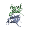
 Assembly
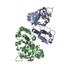


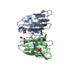
 Mass: 427.201 Da / Num. of mol.: 2 / Source method: obtained synthetically / Formula: C10H15N5O10P2 / Feature type: SUBJECT OF INVESTIGATION / Comment: ADP, energy-carrying molecule*YM
 Mass: 192.124 Da / Num. of mol.: 2 / Source method: obtained synthetically / Formula: C6H8O7
 Mass: 18.015 Da / Num. of mol.: 46 / Source method: isolated from a natural source / Formula: H2O
 Sample preparation
 Processing