 Movie
Movie Controller

[English] 日本語
 Yorodumi
- PDB-1r66: Crystal Structure of DesIV (dTDP-glucose 4,6-dehydratase) from St... -
+ Open data

- Basic information
| Entry | Database: PDB / ID: 1r66 | ||||||
|---|---|---|---|---|---|---|---|
| Title | Crystal Structure of DesIV (dTDP-glucose 4,6-dehydratase) from Streptomyces venezuelae with NAD and TYD bound | ||||||
 Components Components | TDP-glucose-4,6-dehydratase | ||||||
 Keywords Keywords | LYASE / Dehydratase / Rossmann Fold / Short-Chain Dehydrogenase/Reductase | ||||||
| Function / homology |  Function and homology information Function and homology informationdTDP-glucose 4,6-dehydratase / dTDP-glucose 4,6-dehydratase activity / nucleotide-sugar metabolic process / nucleotide binding Similarity search - Function | ||||||
| Biological species |  Streptomyces venezuelae (bacteria) Streptomyces venezuelae (bacteria) | ||||||
| Method |  X-RAY DIFFRACTION / SYNCHROTRON / MOLECULAR REPLACEMENT / Resolution: 1.44 Å X-RAY DIFFRACTION / SYNCHROTRON / MOLECULAR REPLACEMENT / Resolution: 1.44 Å | ||||||
 Authors Authors | Allard, S.T.M. / Cleland, W.W. / Holden, H.M. | ||||||
 Citation Citation | Journal: J.Biol.Chem. / Year: 2004 Title: High Resolution X-ray Structure of dTDP-Glucose 4,6-Dehydratase from Streptomyces venezuelae Authors: Allard, S.T.M. / Cleland, W.W. / Holden, H.M. | ||||||
| History |
|
- Structure visualization
| Structure viewer | Molecule: MolmilJmol/JSmol |
|---|
- Downloads & links
-Download
| PDBx/mmCIF format | 1r66.cif.gz | 85.3 KB | Display | PDBx/mmCIF format |
|---|---|---|---|---|
| PDB format | pdb1r66.ent.gz | 62 KB | Display | PDB format |
| PDBx/mmJSON format | 1r66.json.gz | Tree view | PDBx/mmJSON format | |
| Others |  Other downloads Other downloads |
-Validation report
| Arichive directory | https://data.pdbj.org/pub/pdb/validation_reports/r6/1r66ftp://data.pdbj.org/pub/pdb/validation_reports/r6/1r66 | HTTPS FTP |
|---|
-Related structure data
| Related structure data |  1r6dC  1bxkS C: citing same article ( S: Starting model for refinement |
|---|---|
| Similar structure data |


-Links
 PDBj
PDBj


- Assembly
| Deposited unit | 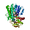
| ||||||||||
|---|---|---|---|---|---|---|---|---|---|---|---|
| 1 | 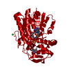
| ||||||||||
| Unit cell |
| ||||||||||
| Components on special symmetry positions |
|
-Components
| #1: Protein | Mass: 36499.887 Da / Num. of mol.: 1 Source method: isolated from a genetically manipulated source Source: (gene. exp.) Streptomyces venezuelae (bacteria) / Gene: DesIV / Plasmid: pET-28 / Production host: |
|---|---|
| #2: Chemical | ChemComp-CL /   Mass: 35.453 Da / Num. of mol.: 1 / Source method: obtained synthetically / Formula: Cl Mass: 35.453 Da / Num. of mol.: 1 / Source method: obtained synthetically / Formula: Cl |
| #3: Chemical | ChemComp-NAD /   Mass: 663.425 Da / Num. of mol.: 1 / Source method: obtained synthetically / Formula: C21H27N7O14P2 / Comment: NAD*YM Mass: 663.425 Da / Num. of mol.: 1 / Source method: obtained synthetically / Formula: C21H27N7O14P2 / Comment: NAD*YM |
| #4: Chemical | ChemComp-TYD /   Mass: 402.188 Da / Num. of mol.: 1 / Source method: obtained synthetically / Formula: C10H16N2O11P2 Mass: 402.188 Da / Num. of mol.: 1 / Source method: obtained synthetically / Formula: C10H16N2O11P2 |
| #5: Water | ChemComp-HOH /  Mass: 18.015 Da / Num. of mol.: 273 / Source method: isolated from a natural source / Formula: H2O Mass: 18.015 Da / Num. of mol.: 273 / Source method: isolated from a natural source / Formula: H2O |
-Experimental details
-Experiment
| Experiment | Method: X-RAY DIFFRACTION / Number of used crystals: 1 |
|---|
- Sample preparation
| Crystal | Density Matthews: 2 Å3/Da / Density % sol: 38.1 % | ||||||||||||||||||||||||||||||||||||||||||||||||
|---|---|---|---|---|---|---|---|---|---|---|---|---|---|---|---|---|---|---|---|---|---|---|---|---|---|---|---|---|---|---|---|---|---|---|---|---|---|---|---|---|---|---|---|---|---|---|---|---|---|
| Crystal grow | Temperature: 277 K / Method: batch / pH: 6.3 Details: PEG 8000, magnesium acetate tetrahydrate, sodium cacodylate, pH 6.3, Batch, temperature 277K | ||||||||||||||||||||||||||||||||||||||||||||||||
| Crystal grow | *PLUS Temperature: 4 ℃ / pH: 6.5 / Method: batch method | ||||||||||||||||||||||||||||||||||||||||||||||||
| Components of the solutions | *PLUS
|
-Data collection
| Diffraction | Mean temperature: 94 K |
|---|---|
| Diffraction source | Source: SYNCHROTRON / Site: APS  / Beamline: 19-BM / Wavelength: 0.9793 Å / Beamline: 19-BM / Wavelength: 0.9793 Å |
| Detector | Type: SBC-3 / Detector: CCD / Date: Dec 4, 2002 / Details: Mirrors |
| Radiation | Monochromator: Si 111 CHANNEL / Protocol: SINGLE WAVELENGTH / Monochromatic (M) / Laue (L): M / Scattering type: x-ray |
| Radiation wavelength | Wavelength: 0.9793 Å / Relative weight: 1 |
| Reflection | Resolution: 1.44→50 Å / Num. all: 57828 / Num. obs: 56518 / % possible obs: 98.6 % / Redundancy: 5.5 % / Rsym value: 0.066 / Net I/σ(I): 26.4 |
| Reflection shell | Resolution: 1.44→1.49 Å / Redundancy: 4 % / Mean I/σ(I) obs: 6.8 / Num. unique all: 5618 / Rsym value: 0.212 / % possible all: 99.9 |
| Reflection | *PLUS Highest resolution: 1.44 Å / Lowest resolution: 50 Å / Rmerge(I) obs: 0.066 |
| Reflection shell | *PLUS % possible obs: 99.9 % / Num. unique obs: 5618 / Rmerge(I) obs: 0.212 |
- Processing
| Software |
| |||||||||||||||||||||||||
|---|---|---|---|---|---|---|---|---|---|---|---|---|---|---|---|---|---|---|---|---|---|---|---|---|---|---|
| Refinement | Method to determine structure: MOLECULAR REPLACEMENT Starting model: PDB ENTRY 1BXK Resolution: 1.44→30 Å / Cross valid method: THROUGHOUT / Stereochemistry target values: Engh & Huber / Details: Used weighted least squares procedure.
| |||||||||||||||||||||||||
| Refinement step | Cycle: LAST / Resolution: 1.44→30 Å
| |||||||||||||||||||||||||
| Refine LS restraints |
| |||||||||||||||||||||||||
| Software | *PLUS Version: 'V. 5-E' / Classification: refinement | |||||||||||||||||||||||||
| Refinement | *PLUS Num. reflection obs: 52099 / Rfactor obs: 0.177 | |||||||||||||||||||||||||
| Solvent computation | *PLUS | |||||||||||||||||||||||||
| Displacement parameters | *PLUS | |||||||||||||||||||||||||
| Refine LS restraints | *PLUS
|
