 Movie
Movie Controller

[English] 日本語
 Yorodumi
- PDB-1r6d: Crystal Structure of DesIV double mutant (dTDP-glucose 4,6-dehydr... -
+ Open data

- Basic information
| Entry | Database: PDB / ID: 1r6d | ||||||
|---|---|---|---|---|---|---|---|
| Title | Crystal Structure of DesIV double mutant (dTDP-glucose 4,6-dehydratase) from Streptomyces venezuelae with NAD and DAU bound | ||||||
 Components Components | TDP-glucose-4,6-dehydratase | ||||||
 Keywords Keywords | LYASE / Dehydratase / Rossmann Fold / Short-Chain Dehydrogenase/Reductase | ||||||
| Function / homology |  Function and homology information Function and homology informationdTDP-glucose 4,6-dehydratase / dTDP-glucose 4,6-dehydratase activity / nucleotide-sugar metabolic process / nucleotide binding Similarity search - Function | ||||||
| Biological species |  Streptomyces venezuelae (bacteria) Streptomyces venezuelae (bacteria) | ||||||
| Method |  X-RAY DIFFRACTION / MOLECULAR REPLACEMENT / Resolution: 1.35 Å X-RAY DIFFRACTION / MOLECULAR REPLACEMENT / Resolution: 1.35 Å | ||||||
 Authors Authors | Allard, S.T.M. / Cleland, W.W. / Holden, H.M. | ||||||
 Citation Citation | Journal: J.Biol.Chem. / Year: 2004 Title: High Resolution X-ray Structure of dTDP-Glucose 4,6-Dehydratase from Streptomyces venezuelae Authors: Allard, S.T.M. / Cleland, W.W. / Holden, H.M. | ||||||
| History |
|
- Structure visualization
| Structure viewer | Molecule: MolmilJmol/JSmol |
|---|
- Downloads & links
-Download
| PDBx/mmCIF format | 1r6d.cif.gz | 88.9 KB | Display | PDBx/mmCIF format |
|---|---|---|---|---|
| PDB format | pdb1r6d.ent.gz | 64.2 KB | Display | PDB format |
| PDBx/mmJSON format | 1r6d.json.gz | Tree view | PDBx/mmJSON format | |
| Others |  Other downloads Other downloads |
-Validation report
| Arichive directory | https://data.pdbj.org/pub/pdb/validation_reports/r6/1r6dftp://data.pdbj.org/pub/pdb/validation_reports/r6/1r6d | HTTPS FTP |
|---|
-Related structure data
| Related structure data | 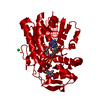 1r66SC S: Starting model for refinement C: citing same article ( |
|---|---|
| Similar structure data |

-Links
 PDBj
PDBj


- Assembly
| Deposited unit | 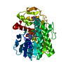
| ||||||||||
|---|---|---|---|---|---|---|---|---|---|---|---|
| 1 | 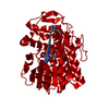
| ||||||||||
| Unit cell |
| ||||||||||
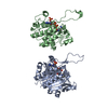
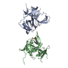
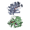
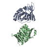
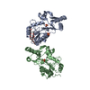
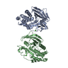
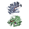
| Details | The biological assembly is a dimer, contructed from chain A (a symmetry partner generated by the two-fold). |
-Components
| #1: Protein | Mass: 36497.918 Da / Num. of mol.: 1 / Mutation: D128N/E129Q Source method: isolated from a genetically manipulated source Source: (gene. exp.) Streptomyces venezuelae (bacteria) / Gene: DesIV / Plasmid: pET-28 / Production host: |
|---|---|
| #2: Chemical | ChemComp-NAD /   Mass: 663.425 Da / Num. of mol.: 1 / Source method: obtained synthetically / Formula: C21H27N7O14P2 / Comment: NAD*YM Mass: 663.425 Da / Num. of mol.: 1 / Source method: obtained synthetically / Formula: C21H27N7O14P2 / Comment: NAD*YM |
| #3: Chemical | ChemComp-DAU / 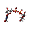  Mass: 564.329 Da / Num. of mol.: 1 / Source method: obtained synthetically / Formula: C16H26N2O16P2 Mass: 564.329 Da / Num. of mol.: 1 / Source method: obtained synthetically / Formula: C16H26N2O16P2 |
| #4: Water | ChemComp-HOH /  Mass: 18.015 Da / Num. of mol.: 371 / Source method: isolated from a natural source / Formula: H2O Mass: 18.015 Da / Num. of mol.: 371 / Source method: isolated from a natural source / Formula: H2O |
-Experimental details
-Experiment
| Experiment | Method: X-RAY DIFFRACTION / Number of used crystals: 3 |
|---|
- Sample preparation
| Crystal | Density Matthews: 1.81 Å3/Da / Density % sol: 31.6 % | ||||||||||||||||||||||||||||||||||||||||||||||||
|---|---|---|---|---|---|---|---|---|---|---|---|---|---|---|---|---|---|---|---|---|---|---|---|---|---|---|---|---|---|---|---|---|---|---|---|---|---|---|---|---|---|---|---|---|---|---|---|---|---|
| Crystal grow | Temperature: 277 K / Method: batch macro-seeding / pH: 6.5 Details: Peg 8000, magnesium chloride, cacodylate, pH 6.5, Batch macro-seeding, temperature 277K | ||||||||||||||||||||||||||||||||||||||||||||||||
| Crystal grow | *PLUS Temperature: 4 ℃ / Method: vapor diffusion, hanging drop | ||||||||||||||||||||||||||||||||||||||||||||||||
| Components of the solutions | *PLUS
|
-Data collection
| Diffraction | Mean temperature: 100 K |
|---|---|
| Diffraction source | Source: ROTATING ANODE / Type: RIGAKU RU200 / Wavelength: 1.5418 Å |
| Detector | Type: SIEMENS HI-STAR / Detector: AREA DETECTOR / Date: Aug 13, 2003 / Details: Gobel focusing optics |
| Radiation | Monochromator: GOEBEL OPTICS / Protocol: SINGLE WAVELENGTH / Monochromatic (M) / Laue (L): M / Scattering type: x-ray |
| Radiation wavelength | Wavelength: 1.5418 Å / Relative weight: 1 |
| Reflection | Resolution: 1.35→30 Å / Num. all: 61664 / Num. obs: 61664 / % possible obs: 91.9 % / Observed criterion σ(F): 0 / Redundancy: 3.6 % / Rsym value: 0.04 / Net I/σ(I): 17.3 |
| Reflection shell | Resolution: 1.35→1.41 Å / Redundancy: 1.7 % / Mean I/σ(I) obs: 2 / Num. unique all: 6573 / Rsym value: 0.223 / % possible all: 81 |
| Reflection | *PLUS Rmerge(I) obs: 0.04 |
| Reflection shell | *PLUS % possible obs: 81 % / Num. unique obs: 6573 / Rmerge(I) obs: 0.223 |
- Processing
| Software |
| |||||||||||||||||||||||||
|---|---|---|---|---|---|---|---|---|---|---|---|---|---|---|---|---|---|---|---|---|---|---|---|---|---|---|
| Refinement | Method to determine structure: MOLECULAR REPLACEMENT Starting model: 1R66 Resolution: 1.35→30 Å / Cross valid method: THROUGHOUT / σ(F): 0 / Stereochemistry target values: Engh & Huber
| |||||||||||||||||||||||||
| Refinement step | Cycle: LAST / Resolution: 1.35→30 Å
| |||||||||||||||||||||||||
| Refine LS restraints |
| |||||||||||||||||||||||||
| Refinement | *PLUS Num. reflection obs: 55477 | |||||||||||||||||||||||||
| Solvent computation | *PLUS | |||||||||||||||||||||||||
| Displacement parameters | *PLUS | |||||||||||||||||||||||||
| Refine LS restraints | *PLUS
|
