 Movie
Movie Controller

[English] 日本語
 Yorodumi
- PDB-4ni3: Crystal Structure of GH29 family alpha-L-fucosidase from Fusarium... -
+ Open data

- Basic information
| Entry | Database: PDB / ID: 4ni3 | |||||||||
|---|---|---|---|---|---|---|---|---|---|---|
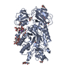
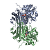
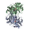
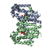
| Title | Crystal Structure of GH29 family alpha-L-fucosidase from Fusarium graminearum in the closed form | |||||||||
 Components Components | Alpha-fucosidase GH29 | |||||||||
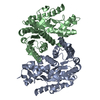
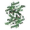
 Keywords Keywords | HYDROLASE / fucosidase / GH29 / glycoside hydrolase / TIM barrel / crystallin | |||||||||
| Function / homology |  Function and homology information Function and homology informationalpha-L-fucosidase / alpha-L-fucosidase activity / fucose metabolic process / glycoside catabolic process Similarity search - Function | |||||||||
| Biological species |  Fusarium graminearum (fungus) Fusarium graminearum (fungus) | |||||||||
| Method |  X-RAY DIFFRACTION / SYNCHROTRON / MOLECULAR REPLACEMENT / Resolution: 1.3993 Å X-RAY DIFFRACTION / SYNCHROTRON / MOLECULAR REPLACEMENT / Resolution: 1.3993 Å | |||||||||
 Authors Authors | Cao, H. / Walton, J.D. / Brumm, P. / Phillips Jr., G.N. | |||||||||
 Citation Citation | Journal: J.Biol.Chem. / Year: 2014 Title: Structure and Substrate Specificity of a Eukaryotic Fucosidase from Fusarium graminearum. Authors: Cao, H. / Walton, J.D. / Brumm, P. / Phillips, G.N. | |||||||||
| History |
|
- Structure visualization
| Structure viewer | Molecule: MolmilJmol/JSmol |
|---|
- Downloads & links
-Download
| PDBx/mmCIF format | 4ni3.cif.gz | 285.9 KB | Display | PDBx/mmCIF format |
|---|---|---|---|---|
| PDB format | pdb4ni3.ent.gz | 226.7 KB | Display | PDB format |
| PDBx/mmJSON format | 4ni3.json.gz | Tree view | PDBx/mmJSON format | |
| Others |  Other downloads Other downloads |
-Validation report
| Arichive directory | https://data.pdbj.org/pub/pdb/validation_reports/ni/4ni3ftp://data.pdbj.org/pub/pdb/validation_reports/ni/4ni3 | HTTPS FTP |
|---|
-Related structure data
| Related structure data |  4pspC 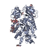 4psrC  1hl8S S: Starting model for refinement C: citing same article ( |
|---|---|
| Similar structure data |


-Links
 PDBj
PDBj
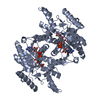
- Assembly
| Deposited unit | 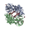
| ||||||||
|---|---|---|---|---|---|---|---|---|---|
| 1 | 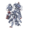
| ||||||||
| 2 | 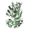
| ||||||||
| Unit cell |
|
-Components
-Protein , 1 types, 2 molecules AB
| #1: Protein | Mass: 65709.781 Da / Num. of mol.: 2 Source method: isolated from a genetically manipulated source Source: (gene. exp.) Fusarium graminearum (fungus) / Gene: FCO1 / Production host: Pichia pastoris (fungus) / References: UniProt: J9UN47 |
|---|
-Sugars , 2 types, 5 molecules 
| #2: Polysaccharide | Source method: isolated from a genetically manipulated source #3: Sugar |  Type: D-saccharide, beta linking / Mass: 221.208 Da / Num. of mol.: 3 Type: D-saccharide, beta linking / Mass: 221.208 Da / Num. of mol.: 3Source method: isolated from a genetically manipulated source Formula: C8H15NO6 |
|---|
-Non-polymers , 4 types, 1418 molecules 


| #4: Chemical | ChemComp-GOL /  Mass: 92.094 Da / Num. of mol.: 8 / Source method: obtained synthetically / Formula: C3H8O3 Mass: 92.094 Da / Num. of mol.: 8 / Source method: obtained synthetically / Formula: C3H8O3#5: Chemical | ChemComp-TRS /  Mass: 122.143 Da / Num. of mol.: 5 / Source method: obtained synthetically / Formula: C4H12NO3 / Comment: pH buffer*YM Mass: 122.143 Da / Num. of mol.: 5 / Source method: obtained synthetically / Formula: C4H12NO3 / Comment: pH buffer*YM#6: Chemical |  Mass: 22.990 Da / Num. of mol.: 3 / Source method: obtained synthetically / Formula: Na Mass: 22.990 Da / Num. of mol.: 3 / Source method: obtained synthetically / Formula: Na#7: Water | ChemComp-HOH / | Mass: 18.015 Da / Num. of mol.: 1402 / Source method: isolated from a natural source / Formula: H2O |
|---|
-Details
| Has protein modification | Y |
|---|
-Experimental details
-Experiment
| Experiment | Method: X-RAY DIFFRACTION / Number of used crystals: 1 |
|---|
- Sample preparation
| Crystal | Density Matthews: 2.08 Å3/Da / Density % sol: 41 % |
|---|---|
| Crystal grow | Temperature: 293.15 K / Method: batch method / pH: 8 Details: 1:1 v/v mixture of 14-16 mg/ml alpha-L-fucosidase (stored in 25mM Tris pH 7.5 and partially deglycosylated by incubation 10:1:1 v/v ratio with EndoH and 500 mM sodium citrate pH 5.5 buffer ...Details: 1:1 v/v mixture of 14-16 mg/ml alpha-L-fucosidase (stored in 25mM Tris pH 7.5 and partially deglycosylated by incubation 10:1:1 v/v ratio with EndoH and 500 mM sodium citrate pH 5.5 buffer from New England Biolabs for more than 24hrs before setting up the drop) with 40% PEG 2000mme, 0.1M Tris pH 8.0, crystals grow within two days, cryoprotected by Mitegen LV cryo-oil, batch method, temperature 293.15K |
-Data collection
| Diffraction | Mean temperature: 100 K |
|---|---|
| Diffraction source | Source: SYNCHROTRON / Site: APS  / Beamline: 21-ID-F / Wavelength: 0.97872 Å / Beamline: 21-ID-F / Wavelength: 0.97872 Å |
| Detector | Type: MARMOSAIC 225 mm CCD / Detector: CCD / Date: Apr 20, 2013 |
| Radiation | Monochromator: Diamond [111] / Protocol: SINGLE WAVELENGTH / Monochromatic (M) / Laue (L): M / Scattering type: x-ray |
| Radiation wavelength | Wavelength: 0.97872 Å / Relative weight: 1 |
| Reflection | Resolution: 1.3993→37.931 Å / Num. all: 209440 / Num. obs: 198748 / % possible obs: 94.9 % / Observed criterion σ(F): 0 / Observed criterion σ(I): 0 / Biso Wilson estimate: 16 Å2 |
| Reflection shell | Resolution: 1.3993→1.42 Å / Redundancy: 3.5 % / Mean I/σ(I) obs: 2.3 / Rsym value: 0.557 / % possible all: 92 |
- Processing
| Software |
| |||||||||||||||||||||||||||||||||||||||||||||||||||||||||||||||||||||||||||||||||||||||||||||||||||||||||
|---|---|---|---|---|---|---|---|---|---|---|---|---|---|---|---|---|---|---|---|---|---|---|---|---|---|---|---|---|---|---|---|---|---|---|---|---|---|---|---|---|---|---|---|---|---|---|---|---|---|---|---|---|---|---|---|---|---|---|---|---|---|---|---|---|---|---|---|---|---|---|---|---|---|---|---|---|---|---|---|---|---|---|---|---|---|---|---|---|---|---|---|---|---|---|---|---|---|---|---|---|---|---|---|---|---|---|
| Refinement | Method to determine structure: MOLECULAR REPLACEMENT Starting model: PDB ENTRY 1HL8 (peptide only) Resolution: 1.3993→37.931 Å / Occupancy max: 1 / Occupancy min: 0.21 / SU ML: 0.13 / σ(F): 1.99 / Phase error: 18.46 / Stereochemistry target values: ML
| |||||||||||||||||||||||||||||||||||||||||||||||||||||||||||||||||||||||||||||||||||||||||||||||||||||||||
| Solvent computation | Shrinkage radii: 0.9 Å / VDW probe radii: 1.11 Å / Solvent model: FLAT BULK SOLVENT MODEL | |||||||||||||||||||||||||||||||||||||||||||||||||||||||||||||||||||||||||||||||||||||||||||||||||||||||||
| Displacement parameters | Biso mean: 21.8 Å2 | |||||||||||||||||||||||||||||||||||||||||||||||||||||||||||||||||||||||||||||||||||||||||||||||||||||||||
| Refinement step | Cycle: LAST / Resolution: 1.3993→37.931 Å
| |||||||||||||||||||||||||||||||||||||||||||||||||||||||||||||||||||||||||||||||||||||||||||||||||||||||||
| Refine LS restraints |
| |||||||||||||||||||||||||||||||||||||||||||||||||||||||||||||||||||||||||||||||||||||||||||||||||||||||||
| LS refinement shell |
|
